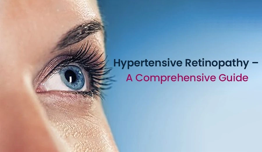
New treatments for hypertensive retinopathy in 2025 include AI imaging, gene therapy, and smart contact lenses. Learn about the latest advances from experts.
Hypertensive retinopathy (HR) is a progressive eye condition caused by prolonged high blood pressure, which damages the delicate blood vessels in the retina. If left untreated, this condition can lead to vision impairment or even permanent blindness.
In recent years, advancements in medical research and technology have introduced new treatments for hypertensive retinopathy, offering more effective management options, early detection tools, and personalized therapeutic approaches.
Table of Contents
In 2025, these innovations are transforming how doctors diagnose, monitor, and treat hypertensive retinopathy. This article explores the latest breakthroughs in treatment, highlighting their benefits and implications for eye health.

Understanding Hypertensive Retinopathy and Its Effects on Vision
Hypertension (high blood pressure) affects millions of people worldwide, and its impact extends beyond heart health. The retina—the light-sensitive tissue at the back of the eye—relies on a healthy blood supply to function properly. Persistent high blood pressure damages these blood vessels, leading to hypertensive retinopathy.
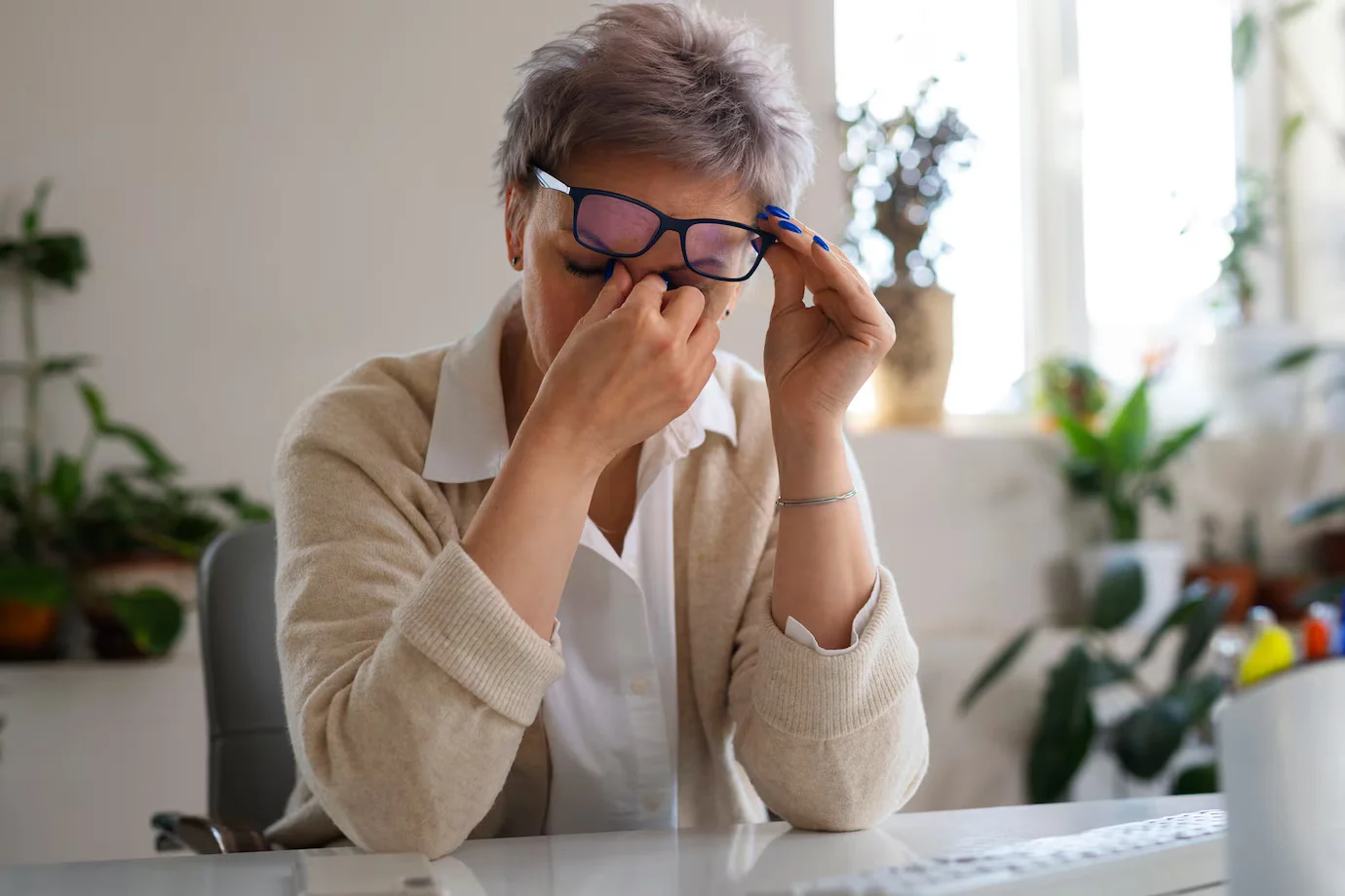
Symptoms of Hypertensive Retinopathy
In the early stages, hypertensive retinopathy may not cause noticeable symptoms. However, as the condition progresses, the following signs may appear:
- Blurry or distorted vision
- Headaches and eye strain
- Reduced night vision
- Double vision or difficulty focusing
- Retinal hemorrhages (bleeding inside the eye)
- Swelling of the optic nerve (severe cases)
How Hypertensive Retinopathy Progresses
Hypertensive retinopathy is categorized into four stages, depending on severity:
- Mild Stage: Slight narrowing of retinal arteries; no symptoms.
- Moderate Stage: Blood vessels become more constricted, causing small hemorrhages.
- Severe Stage: Swelling of the retina and optic nerve; significant vision problems.
- Malignant Hypertensive Retinopathy: A medical emergency that may result in permanent blindness.
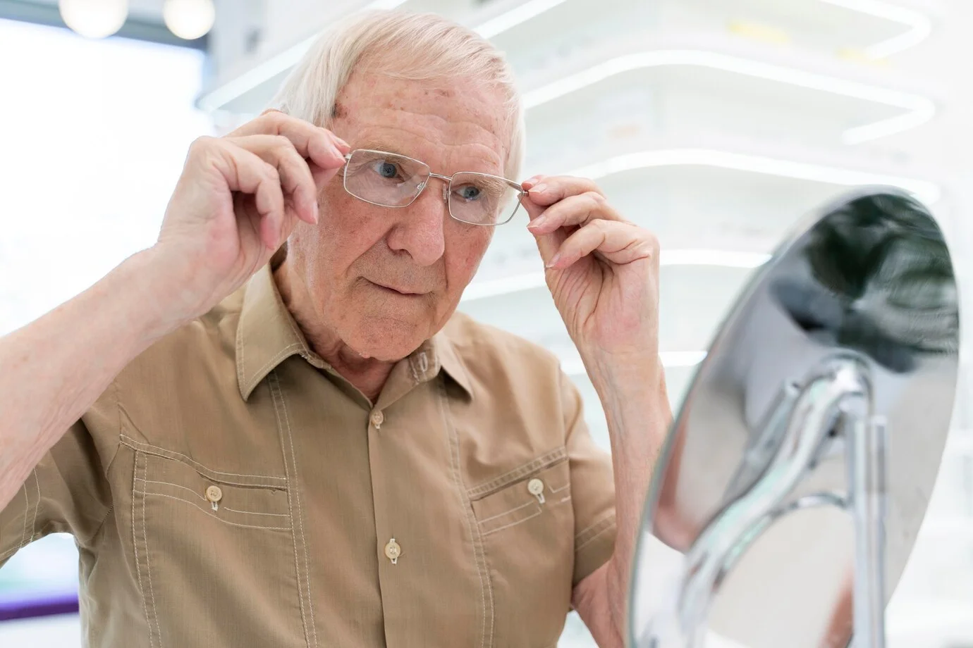
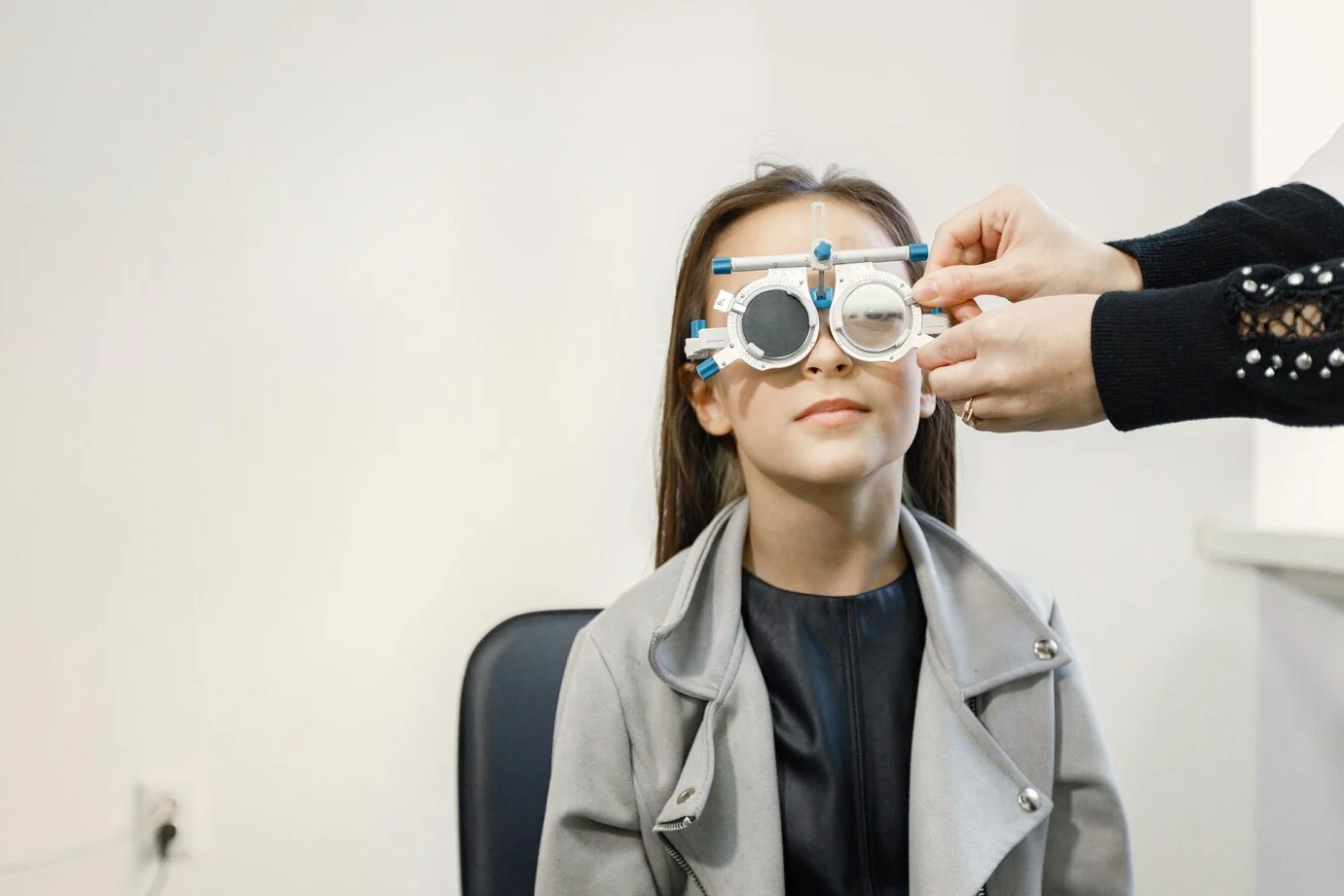
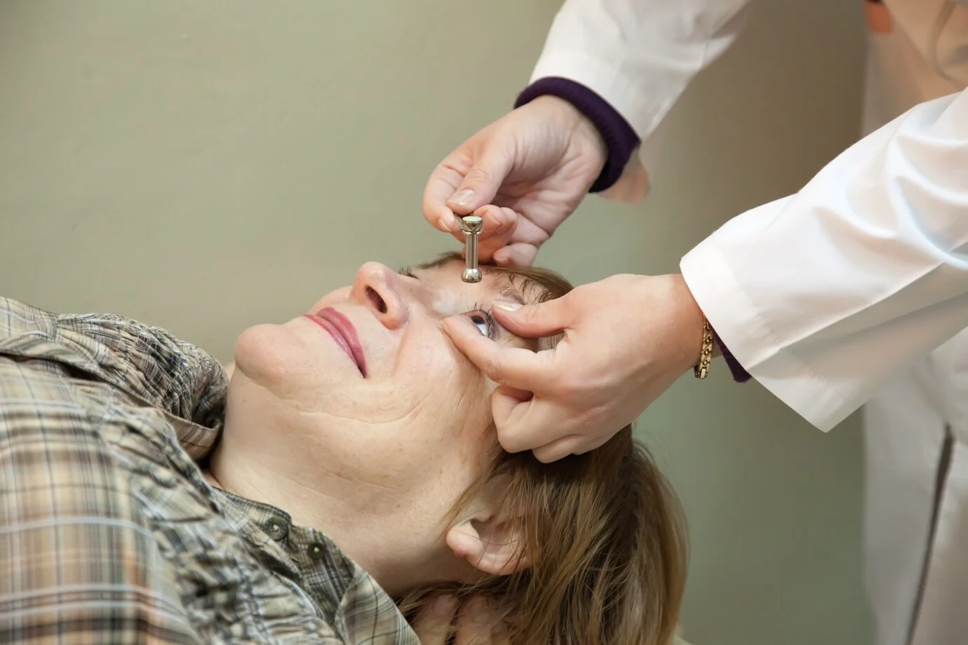
Given the silent nature of early hypertensive retinopathy, routine eye exams are essential for early detection and treatment.
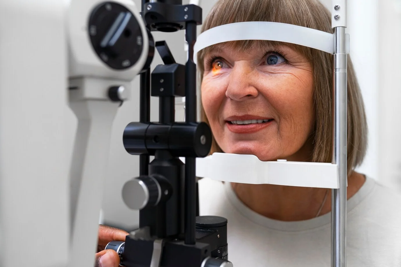
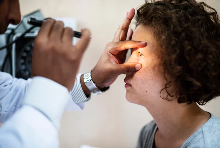
Breakthroughs in Diagnosing Hypertensive Eye Disease
Advancements in medical imaging and AI technology are making it easier to detect Hypertensive Eye Disease at an early stage.
AI-Powered Retinal Screening
Artificial intelligence (AI) has become an essential tool in ophthalmology. AI-driven retinal screening in 2025 offers:
- Faster and more accurate detection of retinal abnormalities.
- Automated detection of microvascular changes in high-risk patients.
- Prediction models that estimate the risk of disease progression.
AI screening improves efficiency and enables early intervention, reducing the risk of severe vision loss.
Ultra-Widefield Imaging
Traditional fundus cameras capture only a portion of the retina. The latest ultra-widefield imaging technology now allows ophthalmologists to:
- View up to 200 degrees of the retina in a single scan.
- Identify early retinal changes before symptoms appear.
- Detect peripheral retinal damage, which may be overlooked in standard exams.
This non-invasive technique ensures quicker and more comprehensive diagnoses.
Optical Coherence Tomography Angiography (OCTA)
OCTA is a revolutionary imaging method that provides a detailed 3D map of retinal blood vessels without the need for contrast dye. This technology allows doctors to:
- Detect early microvascular damage caused by hypertension.
- Monitor blood flow in retinal capillaries with high precision.
- Track disease progression over time to adjust treatments accordingly.
These diagnostic advancements play a crucial role in customizing treatments for Hypertensive Eye Disease
New Treatments for Hypertensive Retinopathy in 2025
The development of new treatments for hypertensive retinopathy is revolutionizing patient care, improving outcomes, and reducing complications.
Gene Therapy – A Revolutionary Approach
Gene therapy is emerging as a potential game-changer for hypertensive retinopathy. Researchers are exploring methods to:
- Modify retinal cells to become resistant to high blood pressure damage.
- Stimulate the repair of damaged blood vessels at a genetic level.
- Reduce oxidative stress and inflammation, which contribute to retinal deterioration.
Although still in experimental stages, gene therapy holds the promise of long-term protection against hypertensive retinopathy.
Personalized Medications for Retinal Protection
Pharmacogenetics—the study of how genes influence drug response—has led to customized medication plans for hypertensive retinopathy patients. These personalized treatments include:
- Retina-protective medications that prevent further vessel damage.
- Targeted anti-inflammatory drugs to slow disease progression.
- Advanced blood pressure management drugs tailored to individual genetic profiles.
By tailoring treatments to each patient’s needs, doctors can minimize side effects and enhance therapeutic effectiveness.
Smart Contact Lenses for Continuous Eye Monitoring
Wearable technology has taken a leap forward with smart contact lenses designed to monitor eye health in real time. These lenses:
- Measure intraocular pressure and retinal blood flow.
- Detect early signs of retinal damage from hypertension.
- Send alerts to doctors or patients when intervention is needed.
This innovation allows for continuous disease management, reducing the risk of sudden vision loss.
Minimally Invasive Laser Treatments
For severe cases, advanced nanosecond laser therapy and microvascular surgery offer safer and more effective treatment options. These include:
- Nanosecond laser therapy to repair retinal blood vessels without damaging surrounding tissue.
- Microvascular bypass surgery to restore normal blood flow in the retina.
- Stem cell therapy to regenerate damaged retinal cells.
With these techniques, patients experience faster recovery times and improved vision preservation.
Preventing Hypertensive Eye Disease: Lifestyle and Medical Tips
While new treatments are promising, prevention remains the most effective strategy. Here’s how to reduce your risk:
Lifestyle Changes
- Maintain a Healthy Blood Pressure: Regular monitoring and medication adherence are crucial.
- Eat a Heart-Healthy Diet: Reduce sodium intake, consume fiber-rich foods, and increase antioxidants.
- Exercise Regularly: Engage in at least 30 minutes of physical activity daily.
- Avoid Smoking and Excessive Alcohol: Both contribute to vascular damage.
- Manage Stress: High stress levels can contribute to hypertension and eye complications.
Medical Recommendations
- Get Regular Eye Checkups: Early detection can prevent severe vision loss.
- Monitor Your Blood Sugar Levels: Hypertension and diabetes often coexist, increasing risk.
- Use Prescription Eye Drops if Recommended: Certain drops can help improve retinal blood flow.
By adopting these habits, individuals can significantly reduce their risk of Hypertensive Eye Disease and protect their vision.

FAQs About New treatments for Hypertensive Retinopathy
1. Can hypertensive retinopathy be cured?
While early stages can be managed with lifestyle changes and medications, advanced cases require medical intervention.
2. What are the newest treatment options for hypertensive retinopathy?
Some of the latest treatments include gene therapy, personalized medications, smart contact lenses, and minimally invasive laser surgery.
3. How often should I get an eye exam if I have high blood pressure?
Patients with hypertension should have an eye exam at least once a year. Those with hypertensive retinopathy may need more frequent checkups.
4. Can I reverse the damage caused by hypertensive retinopathy?
Early-stage damage may improve with blood pressure control, but severe cases often require advanced medical treatments.
5. Where can I get the best treatment for hypertensive retinopathy?
For expert care, specialized eye hospitals offer advanced diagnostic and treatment options.
Ensuring Healthy Vision: The Future of Hypertensive Retinopathy Care
With rapid advancements in new treatments for hypertensive retinopathy, 2025 marks a turning point in eye care. From AI-powered diagnostics to gene therapy and smart wearable devices, patients now have more effective options for managing their condition.
If you or a loved one have high blood pressure, prioritizing regular eye checkups and early intervention at a reputed eye hospital is the key to maintaining healthy vision. Expert care and advanced treatments can help protect your eyesight.