Screen time and kids’ eye health are major concerns in today’s digital world. Learn how excessive screen use affects children’s vision and discover effective ways to protect their eyesight. you can consult best eye hospital in Kolkata regarding this.
Table of Contents

Understanding Screen Time and Its Impact on Kids’ Eye Health
Children today are surrounded by screens—smartphones, tablets, laptops, and televisions. While these devices offer convenience and entertainment, they also pose potential risks to kids’ eye health. The increasing reliance on digital screens for education and leisure has led to a rise in vision-related problems among children.
As a parent, you may wonder: How much screen time is too much? Can screen exposure harm my child’s eyesight? What can I do to minimize the risks? This article explores the connection between screen time and kids’ eye health, along with practical steps to protect your child’s vision.
How Screen Time Affects Kids’ Eye Health
Spending long hours in front of digital devices can lead to various eye-related issues in children. Some of the most common problems include:
1. Digital Eye Strain (Computer Vision Syndrome)
Prolonged screen use can cause digital eye strain, leading to symptoms such as:
- Blurry vision
- Dry or irritated eyes
- Frequent headaches
- Difficulty focusing on distant objects
2. Increased Risk of Myopia (Nearsightedness)
Research suggests that excessive screen exposure, especially at a young age, increases the risk of myopia (nearsightedness). Children who spend more time indoors using screens instead of playing outside are more prone to developing myopia early in life.
3. Disrupted Sleep Patterns
The blue light emitted from screens affects the production of melatonin, a hormone responsible for sleep regulation. Kids who use screens before bedtime often struggle with sleep disturbances, leading to fatigue, irritability, and concentration issues.
4. Reduced Blinking and Dry Eyes
When focusing on screens, kids tend to blink less frequently, which leads to reduced tear production and dry eye syndrome. This can cause discomfort, redness, and itchiness in the eyes.
How Much Screen Time is Too Much?
While digital devices are an inevitable part of modern life, setting healthy screen time limits is essential. According to the American Academy of Pediatrics (AAP), the recommended screen time guidelines for children are:
- Under 18 months – Avoid screen time except for video calls.
- 18 months to 2 years – Introduce only high-quality educational content with parental guidance.
- 2 to 5 years – Limit screen time to one hour per day with educational programs.
- 6 years and above – Maintain consistent screen-free times and encourage outdoor activities.
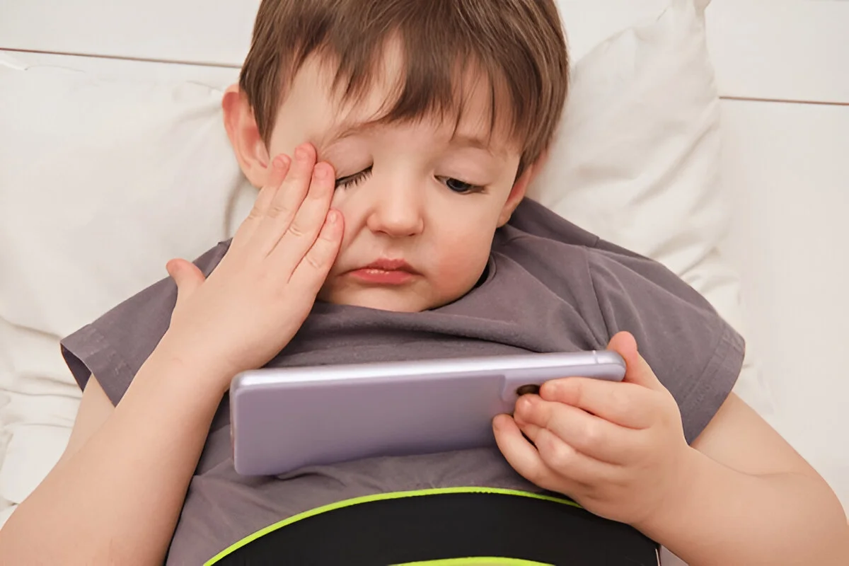
Signs That Your Child May Be Experiencing Eye Strain
If your child frequently experiences any of the following symptoms, it may indicate that screen exposure is affecting their eye health:
- Complaints of blurry vision or double vision
- Frequent rubbing of the eyes
- Increased sensitivity to light
- Difficulty concentrating while reading or studying
- Excessive blinking or watery eyes
If you notice these symptoms, consider reducing screen exposure and consulting an eye specialist for further evaluation.

Tips to Protect Kids’ Eye Health from Excessive Screen Time
To minimize the impact of screen time on kids’ eye health, follow these practical tips:
1. Follow the 20-20-20 Rule
Encourage your child to take a 20-second break every 20 minutes of screen time and look at something 20 feet away. This simple habit helps reduce eye strain.
2. Adjust Screen Brightness and Contrast
- Ensure that screen brightness matches room lighting to avoid excessive glare.
- Use night mode or blue light filters to reduce strain on the eyes, especially in the evening.
3. Maintain a Safe Distance
- Tablets and smartphones should be held at least 18 inches away from the eyes.
- The computer screen should be positioned slightly below eye level to prevent strain.
4. Increase Outdoor Activities
Encourage kids to spend more time playing outdoors, as exposure to natural light helps slow down the progression of myopia.
5. Limit Screen Use Before Bedtime
- Avoid digital screens at least one hour before bedtime to ensure better sleep.
- Introduce bedtime activities like reading books or storytelling instead.
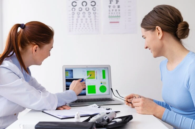
6. Schedule Regular Eye Checkups
Early detection of vision problems can prevent long-term complications. Scheduling routine eye exams at a trusted eye care center ensures your child’s eyesight remains healthy.
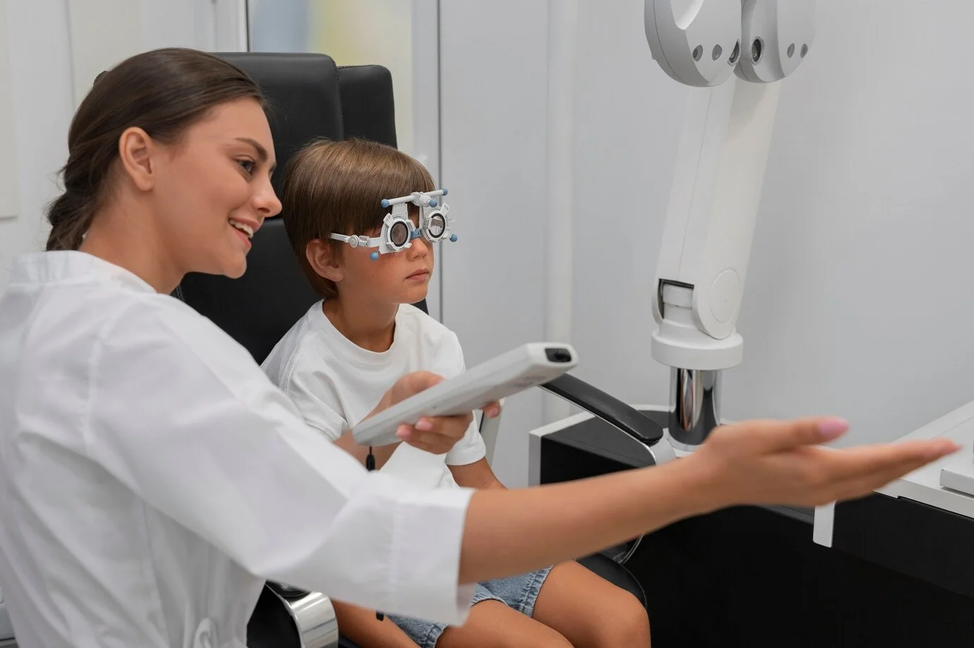
When to Visit an Eye Specialist?
If your child frequently experiences headaches, blurry vision, or difficulty focusing, it’s essential to seek professional help. A comprehensive eye checkup can help detect issues early and provide necessary treatment.
Experienced ophthalmologists specialize in pediatric eye health, providing advanced diagnostic and treatment solutions tailored for children. Regular eye checkups are essential in detecting vision issues early and preventing long-term problems.

Frequently Asked Questions (FAQs)
1. How can I tell if my child’s vision is affected by screen time?
Look out for signs like frequent eye rubbing, complaints of blurry vision, headaches, or difficulty focusing. If symptoms persist, consult an eye specialist for a detailed eye examination.
2. Can blue light glasses help protect my child’s eyes?
Yes, blue light-blocking glasses help reduce eye strain and minimize the impact of blue light exposure, especially during prolonged screen use. However, it’s best to combine them with healthy screen habits.
3. Is screen time worse for younger children?
Yes, excessive screen time at a young age can impact eye development and increase the risk of myopia and eye strain. Controlled and limited screen exposure is essential for younger kids.
4. How often should my child have an eye checkup?
Experts recommend annual eye exams for children, especially if they use screens frequently or show symptoms of vision problems.
5. Does reading on a screen affect eyesight more than reading books?
Yes, reading on a screen for extended periods can cause digital eye strain due to blue light exposure and reduced blinking. Taking breaks and adjusting screen settings can help.
Nurturing Healthy Vision: Small Changes, Big Impact on Eye Health
Screen time and kids’ eye health are closely connected, and excessive exposure to digital devices can lead to various vision-related issues. However, by implementing healthy screen habits, encouraging outdoor activities, and scheduling regular eye checkups, parents can effectively safeguard their child’s eyesight.
Early eye health management is essential for children to prevent long-term vision issues. Regular eye checkups, combined with healthy screen habits and outdoor activities, play a crucial role in maintaining good eyesight. If you’re concerned about your child’s vision, consulting a renowned eye specialist can help detect and address potential problems early.