Prime Eyesight Hospital Near Me Golpark-Gariahat,Kolkata 700029

|

Prime Eyesight Hospital Near Me Baguihati, Vip Road, Kolkata 700059

Best Eye Care Kolkata West Bengal All cashless facilities and West Bengal health scheme available. Top Eye Hospital In Kolkata
Best Eye Care Kolkata

How Can You Solve a Vision Problem with the Best Eye Treatment?

Good vision is vital to perform your daily tasks independently without any struggle. However, many people struggle with serious eye problems such as nearsightedness, astigmatism, cataracts, glaucoma, and farsightedness. Vision problems mainly refer to the condition that damages a person’s clear visibility along with normal vision. Fortunately, with the help of advanced eye care treatment, you can restore and enhance your vision. Among different eye care treatment, cataract treatment in Kolkata is highly beneficial for those who are suffering from cataract. Through this blog, we will discuss various types of eye care treatments that are vital to solving your vision problem.

Types of Vision Problem

In this segment, we will discuss different types of vision problems that many people face during these days-

  1. Farsightedness– It is an eye problem where patient face problem to see nearby objects.
  2. Nearsightedness– In this eye problem, patients face difficulties in seeing distant objects clearly.
  3. Cataracts– This vision problem occurs when the lens in the eye becomes cloudy and slowly causes blurred vision.
  4. Glaucoma– It is serious eye problem that mainly damage the optic nerves and increase the pressure within the eye. This problem leads to peripheral vision loss; if you do not treat it on time, it may lead to permanent blindness.
  5. Dry eye symptoms– In case of insufficient tear production, dry eye problems occur.
  6. Presbyopia-It is an age-related vision problem that people face, and they find difficulties reading books or seeing objects closely.

Best Eye Treatment to Solve Vision Problem

If you want to experience painless eye treatment to solve any of your vision problems, you must visit the best eye hospital in Kolkata. Let’s discuss the different types of eye treatments to solve your vision problems.

  1. Lasik surgery

Lasik surgery reshapes your cornea by using a laser and correcting the vision permanently. Due to its painless eye surgery process, this treatment is in high demand.

  1. Cataract surgery

Cataract surgery mainly removes the cloudy lens and replaces it with an artificially implanted lens. An advanced form of robotic method is available for cataract surgery. The latest technological advancements ensure successful operations without any pain.

  1. Glaucoma treatments

Eye surgeons first use eye drops to reduce intraocular pressure inside the eye before starting a glaucoma operation. Laser therapy or traditional surgical operation methods are available for glaucoma treatment. But before conducting the operation, the eye specialist will examine you and assess your condition.

  1. Use of contact lenses and glass

If you have serious vision problem and cannot see any object from a distances then you must need modern lenses such as anti glare and blue light filters. These options are effective in solving serious ete problems such as hyperopia, astigmatism, and myopia.

How to Choose the Best Eye Treatment Institute

If you are searching online for the best eye treatment institute, you must consider a few factors; let’s discuss-

  1. Eye care institutes must have highly skilled eye surgeons who are famous for their expertise.
  2. The institute must use the latest technology to treat the patients without any hassle.
  3. A personalized treatment plan is also vital for patients because each patient has a unique health condition. Thus, eye surgeons must give special care to each patient to enhance the surgical experience.
  4. The eye care institute must have a good reputation in the market regarding the quality of treatment and cost of the treatment.

Conclusion

Vision problems can be a great problem, but if you choose the best eye care treatment and best eye care institute, you can easily solve any eye-related problem. At Pushpanjali Eye Care Clinic, you will get the best eye treatment with a personalized treatment facility. Our highly skilled and experienced eye surgeons perform their tasks with great intensity.

Squint Eye Myths and Facts: What You Need to Know

Hello Reader!

Welcome to the blog page of Pushpanjali Eye Care, one of the best eye hospital in Kolkata. 

Squint eye, also known as strabismus, is a condition where the eyes do not align properly and look in different directions. This eye disorder can affect people of all ages and may lead to vision impairment if left untreated. However, there are numerous myths surrounding squint eye that often create unnecessary fear and misconceptions. At Pushpanjali Eye Care, the best eye hospital in Kolkata, we aim to clear up these misconceptions and provide the right information.

Table of Contents

Squint eye, Best Eye Hospital In Kolkata , Best ophthalmologist in Kolkata , Best Eye specialists in Kolkata , Top eye hospital in Kolkata , best Glaucoma treatment in Kolkata , Best Lasik surgery in Kolkata , Best Cataract Treatment in Kolkata , best cataract surgery in Kolkata , Lasik Surgery Cost in Kolkata , Cataract surgery in Kolkata , Cataract surgery cost in Kolkata , Glaucoma specialist in Kolkata , Refractive surgery in Kolkata , Lasik surgery in Kolkata

Myth 1: Squint Eye is Just a Cosmetic Issue

Fact: Squint Eye is a Medical Condition That Needs Treatment

Many believe that squint eye is only a cosmetic concern and does not impact vision. However, this is far from the truth. Squint eye can lead to double vision, lazy eye (amblyopia), and even loss of depth perception. Early diagnosis and proper treatment at a reputed facility like Pushpanjali Eye Care, the best eye hospital in Kolkata, can help in managing and correcting this condition effectively.

Myth 2: Squint Eye Will Resolve on Its Own with Age

Fact: Squint Eye Often Requires Medical Intervention

One common misconception is that children with squint eye will eventually outgrow the condition. While some mild cases may improve naturally, most require early intervention, corrective lenses, vision therapy, or surgery to prevent long-term complications. If your child has squint eye, visiting the best eye hospital in Kolkata for early assessment is crucial.

Myth 3: Squint Eye is Always Present from Birth

Fact: Squint Eye Can Develop at Any Age

While many cases of squint eye are congenital, this condition can also develop in adults due to nerve disorders, trauma, diabetes, or other underlying health issues. Sudden onset of squint in adults should not be ignored, as it may indicate an underlying neurological condition. Pushpanjali Eye Care provides expert diagnosis and personalized treatment plans for patients of all ages.

Myth 4: Squint Eye Can Be Cured with Home Remedies

Fact: Squint Eye Requires Professional Medical Treatment

Some people believe that eye exercises, herbal remedies, or wearing black beads can correct squint eye. Unfortunately, these methods are not scientifically proven. Squint eye should be evaluated and treated by an ophthalmologist at a reputed facility like Pushpanjali Eye Care, the best eye hospital in Kolkata, to ensure effective and safe treatment options.

Squint eye, Best Eye Hospital In Kolkata , Best ophthalmologist in Kolkata , Best Eye specialists in Kolkata , Top eye hospital in Kolkata , best Glaucoma treatment in Kolkata , Best Lasik surgery in Kolkata , Best Cataract Treatment in Kolkata , best cataract surgery in Kolkata , Lasik Surgery Cost in Kolkata , Cataract surgery in Kolkata , Cataract surgery cost in Kolkata , Glaucoma specialist in Kolkata , Refractive surgery in Kolkata , Lasik surgery in Kolkata

Myth 5: Squint Surgery is Risky and Ineffective

Fact: Squint Surgery is Safe and Highly Effective

There is a misconception that surgery for squint eye is dangerous or ineffective. In reality, squint eye surgery is a well-established and safe procedure performed under expert care. At Pushpanjali Eye Care, our team of specialists ensures the highest success rates, helping patients regain proper eye alignment and improved vision.

Myth 6: Squint Eye Cannot Be Treated After Childhood

Fact: Adults with Squint Eye Can Also Benefit from Treatment

Although early intervention is ideal, adults with squint eye can still undergo treatment to improve eye alignment and vision. Advanced treatments, including prism glasses, Botox injections, and corrective surgery, can significantly enhance the quality of life for adult patients.

Myth 7: Wearing Glasses for Squint Eye is Useless

Fact: Glasses Can Help in Many Cases

Some types of squint eye, especially those caused by refractive errors, can be corrected with prescription glasses. Pushpanjali Eye Care offers a comprehensive eye examination to determine the best corrective solution for each patient.

Squint eye, Best Eye Hospital In Kolkata , Best ophthalmologist in Kolkata , Best Eye specialists in Kolkata , Top eye hospital in Kolkata , best Glaucoma treatment in Kolkata , Best Lasik surgery in Kolkata , Best Cataract Treatment in Kolkata , best cataract surgery in Kolkata , Lasik Surgery Cost in Kolkata , Cataract surgery in Kolkata , Cataract surgery cost in Kolkata , Glaucoma specialist in Kolkata , Refractive surgery in Kolkata , Lasik surgery in Kolkata

Can squint eyes affect a child’s self-confidence?

Yes, squint eyes can significantly affect a child’s self-confidence. Children with squint eyes often face emotional challenges due to their appearance, which can lead to feelings of embarrassment and self-consciousness. This can have a lasting impact on their self-esteem and confidence, particularly as they grow older and become more aware of their condition.

The effects of squint eyes on a child’s self-confidence can manifest in several ways:

  1. Social interactions: Children with squint eyes may find it difficult to engage in typical social interactions, avoiding eye contact or feeling uncomfortable in group settings. This can hinder their ability to form and maintain friendships, potentially leading to social isolation.
  2. Bullying: Unfortunately, children with visible squint eyes may face teasing or bullying from peers, which can exacerbate their self-esteem issues.
  3. Academic performance: The constant struggle with their visual condition can affect a child’s academic performance and participation in activities, further impacting their overall emotional well-being.
  4. Appearance concerns: As children grow older, they may become increasingly self-conscious about their appearance, which can affect their self-confidence and social interactions.

It’s important to note that early intervention and treatment can help mitigate these effects. Parents and caregivers play a crucial role in supporting children with squint eyes by providing a supportive environment, encouraging a positive self-image, and seeking timely medical treatment or therapy.

Are there any non-surgical treatments for squint eyes?

Yes, there are several effective non-surgical treatments for squint eyes (strabismus) that can help improve eye alignment and coordination. Here are the primary options:

1. Prescription Glasses

Many squint cases are associated with refractive errors such as nearsightedness, farsightedness, or astigmatism. Corrective lenses can help alleviate these issues, improving overall eye alignment and function.

2. Vision Therapy

Vision therapy consists of a series of customized exercises designed to strengthen eye muscles and improve coordination. This therapy is particularly effective for children and adults with mild to moderate squints. Exercises may include:

  • Eye Tracking: Following moving objects to enhance coordination.
  • Pencil Push-ups: Focusing on a pencil as it is moved closer to the nose, helping improve convergence.

3. Eye Patching

This method involves covering the stronger eye with a patch to encourage the weaker eye to work harder, promoting better coordination and strengthening of the visual pathways. Patching is especially beneficial for children.

4. Prism Lenses

Prism lenses can be incorporated into regular eyeglasses to help redirect light entering the eye, which aids in aligning the visual input from both eyes. This non-invasive option is useful for correcting double vision associated with squints.

5. Botox Injections

In some cases, botulinum toxin (Botox) injections can be used to temporarily weaken overactive eye muscles, allowing for better alignment of the eyes. This method is less invasive than surgery and can provide significant improvement in certain types of squints.

Also Read: Why Your Vision Blurs After Too Much Screen Time – And How to Fix It?

Squint eye, Best Eye Hospital In Kolkata , Best ophthalmologist in Kolkata , Best Eye specialists in Kolkata , Top eye hospital in Kolkata , best Glaucoma treatment in Kolkata , Best Lasik surgery in Kolkata , Best Cataract Treatment in Kolkata , best cataract surgery in Kolkata , Lasik Surgery Cost in Kolkata , Cataract surgery in Kolkata , Cataract surgery cost in Kolkata , Glaucoma specialist in Kolkata , Refractive surgery in Kolkata , Lasik surgery in Kolkata

Yes, there are several specific exercises recommended for adults with squint eyes (strabismus) that can help improve eye alignment and coordination. Here are some effective exercises:

1. Pencil Push-Ups

This exercise helps both eyes focus on a single object, which is essential for improving coordination.

  • Hold a pencil at arm’s length, pointing it away from you.
  • Focus on a letter or number on the pencil.
  • Slowly bring the pencil closer to your nose while maintaining focus.
  • Stop when your vision becomes blurry, then repeat the process.

2. Brock String

This exercise enhances eye coordination and is particularly useful for crossed eyes.

  • Take a string about five feet long and attach one end to a fixed point.
  • Thread three different colored beads onto the string, spaced apart.
  • Hold the other end of the string against your nose.
  • Focus on each bead in turn; you should see an “X” where the strings cross at each bead. If not, your eyes may not be properly aligned.

3. Focus Shifting

This exercise improves depth perception and eye muscle flexibility.

  • Choose two objects: one close (like a book) and one far away (like a picture on the wall).
  • Shift your gaze from the close object to the far one and back again.
  • Repeat this process slowly to enhance overall eye alignment.

4. Eye Tracking

This exercise helps increase muscle control and coordination.

  • Hold a pen or your finger in front of your face.
  • Move it horizontally (left to right) and vertically (up and down).
  • Follow the movement with your eyes without moving your head.

5. Cover-Uncover Exercise

This exercise trains each eye individually, which can help improve strength and alignment.

  • Cover one eye with your hand and focus on an object with the uncovered eye for a few seconds.
  • Switch and repeat with the other eye.

6. Near-Far Focus

This technique helps strengthen eye muscles by alternating focus between near and far objects.

  • Hold an object close to your eyes, focusing on it.
  • Then shift your gaze to a distant object.
  • Alternate between near and far objects several times.
Squint eye, Best Eye Hospital In Kolkata , Best ophthalmologist in Kolkata , Best Eye specialists in Kolkata , Top eye hospital in Kolkata , best Glaucoma treatment in Kolkata , Best Lasik surgery in Kolkata , Best Cataract Treatment in Kolkata , best cataract surgery in Kolkata , Lasik Surgery Cost in Kolkata , Cataract surgery in Kolkata , Cataract surgery cost in Kolkata , Glaucoma specialist in Kolkata , Refractive surgery in Kolkata , Lasik surgery in Kolkata

How does vision therapy help in correcting squint?

Vision therapy is a non-surgical treatment option for correcting squint eyes (strabismus) that aims to improve eye coordination and strengthen the eye muscles. Here’s how vision therapy helps in addressing squint eyes:

1. Customized Exercise Programs

Vision therapy involves a series of tailored exercises designed to meet the specific needs of the patient. These exercises focus on enhancing eye alignment, improving binocular vision, and helping both eyes work together more effectively. Regular sessions with a trained optometrist or ophthalmologist allow patients to engage in activities that promote muscle strength and coordination.

2. Strengthening Eye Muscles

Through targeted exercises, vision therapy strengthens the muscles that control eye movement. This is crucial for individuals with squint eyes, as weak or uncoordinated muscles can lead to misalignment. By practicing these exercises consistently, patients can improve their muscle control and reduce the degree of squint.

3. Enhancing Coordination

Vision therapy helps train the brain to better control eye movements, improving coordination between the two eyes. This is particularly important for individuals with squint eyes, as proper coordination is essential for achieving clear and single vision. Exercises may include tracking moving objects, focusing on near and far targets, and using tools like prisms to enhance visual perception.

4. Improving Binocular Vision

One of the primary goals of vision therapy is to enhance binocular vision—the ability to use both eyes together effectively. This is vital for depth perception and overall visual function. By engaging in activities that promote teamwork between the eyes, patients can work towards achieving better alignment and visual harmony.

5. Addressing Underlying Issues

In some cases, squint eyes may be associated with underlying refractive errors or other visual issues. Vision therapy can help address these problems by incorporating corrective lenses or other interventions into the treatment plan, ensuring a comprehensive approach to improving vision.

6. Monitoring Progress

Regular follow-ups with an eye care specialist during vision therapy allow for ongoing assessment of progress. Adjustments can be made to the exercise regimen based on individual improvement, ensuring that each patient receives personalized care tailored to their specific condition.

Squint eye, Best Eye Hospital In Kolkata , Best ophthalmologist in Kolkata , Best Eye specialists in Kolkata , Top eye hospital in Kolkata , best Glaucoma treatment in Kolkata , Best Lasik surgery in Kolkata , Best Cataract Treatment in Kolkata , best cataract surgery in Kolkata , Lasik Surgery Cost in Kolkata , Cataract surgery in Kolkata , Cataract surgery cost in Kolkata , Glaucoma specialist in Kolkata , Refractive surgery in Kolkata , Lasik surgery in Kolkata

FAQ

1. Can squint eye be treated without surgery?

Yes, in many cases, squint eye can be managed with glasses, vision therapy, or Botox injections. Surgery is recommended only for severe cases.

2. Is squint eye hereditary?

Squint eye can have a genetic component, meaning it may run in families. However, environmental and neurological factors also play a role.

3. At what age should squint eye be treated?

Early intervention is best. Treatment should ideally begin in childhood, but adults can also benefit from corrective procedures.

4. Does squint eye cause blindness?

While squint eye itself does not cause blindness, untreated cases can lead to amblyopia (lazy eye), which can significantly reduce vision in one eye.

5. Can an adult develop squint eye suddenly?

Yes, sudden squint eye in adults can be due to nerve damage, stroke, or other medical conditions and should be evaluated by an eye specialist immediately.

Conclusion

Squint eye is a condition that requires medical attention, and believing in myths can delay effective treatment. If you or your child has squint eye, seeking professional care at Pushpanjali Eye Care, the best eye hospital in Kolkata, is the right step toward better vision and quality of life. Our expert ophthalmologists are here to guide you through the best treatment options and ensure optimal eye health.

Book an appointment with Pushpanjali Eye Care today to get the right diagnosis and treatment for squint eye!

10 Signs You Need an Eye Exam Which Most People Ignore

A routine Eye Exam is crucial for detecting hidden vision issues. With professional eye care, you can identify early warning signs before they become serious problems. Learn the top 10 overlooked signs that indicate it’s time for an eye check-up and stay ahead of vision concerns!

10 Silent Signs You Need an Eye Exam – Don’t Ignore Them!

Our eyes are one of the most vital organs, yet they are often neglected when it comes to regular health check-ups. Most people assume that unless they have blurry vision, they don’t need an eye exam. However, many subtle signs indicate underlying issues that, if left untreated, can lead to severe vision problems.

Eye Exam

Whether it’s frequent headaches, difficulty reading, or dry eyes, these symptoms may suggest something more serious. This guide will walk you through 10 commonly ignored signs that indicate it’s time for an eye check-up—helping you take charge of your vision before it’s too late.

1. Frequent Headaches

If you find yourself reaching for painkillers due to recurring headaches, it might not just be stress or dehydration—your eyes could be the culprit. Uncorrected vision issues, eye strain, or even astigmatism can put undue pressure on the eyes, leading to persistent headaches. If this is a regular occurrence, scheduling an eye exam is the best way to determine the root cause.

2. Struggling to See at Night

Do you find it hard to drive at night because of glare from headlights or poor visibility? Difficulty seeing in low-light conditions is an early sign of night blindness (nyctalopia). This could be caused by a vitamin A deficiency, cataracts, or even early-stage glaucoma. A comprehensive eye check-up can help diagnose the problem early.

3. Frequent Eye Strain and Fatigue

In today’s digital world, spending hours on screens is common, leading to digital eye strain. Symptoms like burning, itching, or heavy eyelids after prolonged screen use could mean your eyes are overworked. While some rest may help, persistent strain indicates a need for a professional evaluation.

How to Prevent Digital Eye Strain?

  • Follow the 20-20-20 rule: Every 20 minutes, look at something 20 feet away for 20 seconds.
  • Adjust screen brightness to match your surroundings.
  • Blink frequently to prevent dryness.

4. Blurry or Double Vision

Sudden or progressive blurry vision should never be ignored. It could signal refractive errors, cataracts, or even neurological issues. Double vision (diplopia), on the other hand, may indicate misalignment of the eyes or muscle problems that need urgent attention.

5. Seeing Floaters or Flashing Lights

Tiny floating specks in your vision (floaters) are usually harmless, but if you notice a sudden increase in floaters, flashes of light, or a shadowy curtain over your vision, it could be a sign of retinal detachment—a condition that requires immediate medical care to prevent vision loss.

6. Difficulty Reading or Focusing on Close Objects

Do you struggle to read books, menus, or messages on your phone? If holding things farther away helps you focus, you may have presbyopia, a common condition as we age. It usually starts in the early 40s and requires corrective lenses to improve near vision.

7. Sensitivity to Light (Photophobia)

Unusual light sensitivity can be a symptom of various eye conditions like corneal abrasions, infections, or even migraines. If bright lights make you squint or cause discomfort, consult an eye specialist for a thorough evaluation.

8. Dry, Itchy, or Watery Eyes

Chronic dry eyes can be more than just an irritation—it could indicate dry eye syndrome, which occurs when your eyes fail to produce enough tears. Conversely, excessively watery eyes might be a response to dryness or an underlying allergy. An eye test can help determine the cause and suggest appropriate treatment.

9. Trouble Adapting to Light Changes

If your eyes take too long to adjust when moving from a bright environment to a dark one (or vice versa), it could signal aging-related vision problems, cataracts, or retinal disorders. A professional eye check-up can detect these conditions early.

10. Frequent Eye Infections or Redness

Persistent redness, swelling, or irritation could indicate conditions like conjunctivitis, blepharitis, or even an allergic reaction. If eye infections keep coming back, it might be a sign of an underlying problem that requires professional diagnosis.

Why Regular Eye Exams Are Essential

Even if you don’t experience any of these symptoms, scheduling regular eye exams is crucial to maintain long-term eye health. Early detection of conditions like glaucoma, diabetic retinopathy, and macular degeneration can prevent permanent vision loss.

How Often Should You Get an Eye Exam?

  • Children: Once every 1-2 years
  • Adults (18-40): Every 2 years
  • Adults (40+): Annually, or as recommended by an eye doctor

If you have diabetes, hypertension, or a family history of eye diseases, more frequent eye exams may be necessary.

FAQs

1. What age should I start getting regular eye exams?

Eye check-ups should start from childhood, but adults over 40 should have more frequent exams to monitor age-related vision changes.

2. Can an eye exam detect other health conditions?

Yes! Diabetes, high blood pressure, and even brain tumors can sometimes be detected through an eye exam before other symptoms appear.

3. How long does an eye exam take?

A standard eye check-up typically takes 30-60 minutes, depending on the tests required.

4. Is blurry vision always a sign of poor eyesight?

Not necessarily. Blurry vision can be caused by dry eyes, fatigue, or underlying conditions like cataracts. Only an eye doctor can provide a definite diagnosis.

5. Can prolonged screen use permanently damage eyesight?

While digital screens don’t cause permanent damage, excessive screen time can lead to eye strain, dry eyes, and discomfort. Proper screen habits help minimize these effects.

Don’t Overlook the Signs – Prioritize Your Eye Health Today!

Ignoring early signs of vision problems can lead to complications that may affect your daily life. Whether it’s frequent headaches, blurry vision, or difficulty seeing at night, an eye exam can diagnose issues early and prevent future complications.

For those seeking expert eye care, comprehensive eye check-ups with state-of-the-art facilities and experienced professionals can make a significant difference. Prioritize your vision today—because clear sight means a brighter future!

Screen Time and Kids’ Eye Health: What Parents Should Know

Screen time and kids’ eye health are major concerns in today’s digital world. Learn how excessive screen use affects children’s vision and discover effective ways to protect their eyesight. you can consult best eye hospital in Kolkata regarding this.

Understanding Screen Time and Its Impact on Kids’ Eye Health

Children today are surrounded by screens—smartphones, tablets, laptops, and televisions. While these devices offer convenience and entertainment, they also pose potential risks to kids’ eye health. The increasing reliance on digital screens for education and leisure has led to a rise in vision-related problems among children.

As a parent, you may wonder: How much screen time is too much? Can screen exposure harm my child’s eyesight? What can I do to minimize the risks? This article explores the connection between screen time and kids’ eye health, along with practical steps to protect your child’s vision.

How Screen Time Affects Kids’ Eye Health

Spending long hours in front of digital devices can lead to various eye-related issues in children. Some of the most common problems include:

1. Digital Eye Strain (Computer Vision Syndrome)

Prolonged screen use can cause digital eye strain, leading to symptoms such as:

  • Blurry vision
  • Dry or irritated eyes
  • Frequent headaches
  • Difficulty focusing on distant objects

2. Increased Risk of Myopia (Nearsightedness)

Research suggests that excessive screen exposure, especially at a young age, increases the risk of myopia (nearsightedness). Children who spend more time indoors using screens instead of playing outside are more prone to developing myopia early in life.

3. Disrupted Sleep Patterns

The blue light emitted from screens affects the production of melatonin, a hormone responsible for sleep regulation. Kids who use screens before bedtime often struggle with sleep disturbances, leading to fatigue, irritability, and concentration issues.

4. Reduced Blinking and Dry Eyes

When focusing on screens, kids tend to blink less frequently, which leads to reduced tear production and dry eye syndrome. This can cause discomfort, redness, and itchiness in the eyes.

How Much Screen Time is Too Much?

While digital devices are an inevitable part of modern life, setting healthy screen time limits is essential. According to the American Academy of Pediatrics (AAP), the recommended screen time guidelines for children are:

  • Under 18 months – Avoid screen time except for video calls.
  • 18 months to 2 years – Introduce only high-quality educational content with parental guidance.
  • 2 to 5 years – Limit screen time to one hour per day with educational programs.
  • 6 years and above – Maintain consistent screen-free times and encourage outdoor activities.

Signs That Your Child May Be Experiencing Eye Strain

If your child frequently experiences any of the following symptoms, it may indicate that screen exposure is affecting their eye health:

  • Complaints of blurry vision or double vision
  • Frequent rubbing of the eyes
  • Increased sensitivity to light
  • Difficulty concentrating while reading or studying
  • Excessive blinking or watery eyes

If you notice these symptoms, consider reducing screen exposure and consulting an eye specialist for further evaluation.

Tips to Protect Kids’ Eye Health from Excessive Screen Time

To minimize the impact of screen time on kids’ eye health, follow these practical tips:

1. Follow the 20-20-20 Rule

Encourage your child to take a 20-second break every 20 minutes of screen time and look at something 20 feet away. This simple habit helps reduce eye strain.

2. Adjust Screen Brightness and Contrast

  • Ensure that screen brightness matches room lighting to avoid excessive glare.
  • Use night mode or blue light filters to reduce strain on the eyes, especially in the evening.

3. Maintain a Safe Distance

  • Tablets and smartphones should be held at least 18 inches away from the eyes.
  • The computer screen should be positioned slightly below eye level to prevent strain.

4. Increase Outdoor Activities

Encourage kids to spend more time playing outdoors, as exposure to natural light helps slow down the progression of myopia.

5. Limit Screen Use Before Bedtime

  • Avoid digital screens at least one hour before bedtime to ensure better sleep.
  • Introduce bedtime activities like reading books or storytelling instead.

6. Schedule Regular Eye Checkups

Early detection of vision problems can prevent long-term complications. Scheduling routine eye exams at a trusted eye care center ensures your child’s eyesight remains healthy.

When to Visit an Eye Specialist?

If your child frequently experiences headaches, blurry vision, or difficulty focusing, it’s essential to seek professional help. A comprehensive eye checkup can help detect issues early and provide necessary treatment.

Experienced ophthalmologists specialize in pediatric eye health, providing advanced diagnostic and treatment solutions tailored for children. Regular eye checkups are essential in detecting vision issues early and preventing long-term problems.

Frequently Asked Questions (FAQs)

1. How can I tell if my child’s vision is affected by screen time?

Look out for signs like frequent eye rubbing, complaints of blurry vision, headaches, or difficulty focusing. If symptoms persist, consult an eye specialist for a detailed eye examination.

2. Can blue light glasses help protect my child’s eyes?

Yes, blue light-blocking glasses help reduce eye strain and minimize the impact of blue light exposure, especially during prolonged screen use. However, it’s best to combine them with healthy screen habits.

3. Is screen time worse for younger children?

Yes, excessive screen time at a young age can impact eye development and increase the risk of myopia and eye strain. Controlled and limited screen exposure is essential for younger kids.

4. How often should my child have an eye checkup?

Experts recommend annual eye exams for children, especially if they use screens frequently or show symptoms of vision problems.

5. Does reading on a screen affect eyesight more than reading books?

Yes, reading on a screen for extended periods can cause digital eye strain due to blue light exposure and reduced blinking. Taking breaks and adjusting screen settings can help.

Nurturing Healthy Vision: Small Changes, Big Impact on Eye Health

Screen time and kids’ eye health are closely connected, and excessive exposure to digital devices can lead to various vision-related issues. However, by implementing healthy screen habits, encouraging outdoor activities, and scheduling regular eye checkups, parents can effectively safeguard their child’s eyesight.

Early eye health management is essential for children to prevent long-term vision issues. Regular eye checkups, combined with healthy screen habits and outdoor activities, play a crucial role in maintaining good eyesight. If you’re concerned about your child’s vision, consulting a renowned eye specialist can help detect and address potential problems early.

Why Your Vision Blurs After Too Much Screen Time – And How to Fix It?

Hello Reader!

Welcome to the blog page of Pushpanjali Eye Care, one of the best eye hospital in Kolkata. 

In today’s digital age, our reliance on screens has significantly increased. Whether it’s for work, entertainment, or social interaction, we spend hours staring at computers, smartphones, and tablets. However, too much screen time often leads to vision blur, eye strain, and discomfort. If you frequently experience blurred vision after prolonged screen use, it’s essential to understand the causes and solutions to maintain optimal eye health. As the best eye hospital in Kolkata, Pushpanjali Eye Care is here to guide you on why this happens and how to fix it.

vision blur, Best Eye Hospital In Kolkata , Best ophthalmologist in Kolkata , Best Eye specialists in Kolkata , Top eye hospital in Kolkata , best Glaucoma treatment in Kolkata , Best Lasik surgery in Kolkata , Best Cataract Treatment in Kolkata , best cataract surgery in Kolkata , Lasik Surgery Cost in Kolkata , Cataract surgery in Kolkata , Cataract surgery cost in Kolkata , Glaucoma specialist in Kolkata , Refractive surgery in Kolkata , Lasik surgery in Kolkata

Why Does Too Much Screen Time Cause Vision Blur?

1. Digital Eye Strain (Computer Vision Syndrome)

Spending long hours in front of screens can cause Digital Eye Strain, also known as Computer Vision Syndrome (CVS). The constant focus on the screen without breaks leads to fatigue in the eye muscles, resulting in vision blur.

2. Reduced Blinking

Normally, we blink about 15-20 times per minute. However, while staring at screens, the blink rate reduces significantly. This leads to dry eyes, irritation, and temporary vision blur.

3. Blue Light Exposure

Digital screens emit blue light, which can disrupt our natural sleep-wake cycle and strain the eyes. Prolonged exposure to blue light can contribute to vision blur and discomfort.

4. Poor Screen Positioning

Incorrect screen placement, such as holding your phone too close or positioning your computer too high, forces your eyes to overwork, causing strain and vision blur.

5. Underlying Vision Problems

Too much screen time can exacerbate existing eye conditions like myopia (nearsightedness) or hyperopia (farsightedness), making vision blur more noticeable.

vision blur, Best Eye Hospital In Kolkata , Best ophthalmologist in Kolkata , Best Eye specialists in Kolkata , Top eye hospital in Kolkata , best Glaucoma treatment in Kolkata , Best Lasik surgery in Kolkata , Best Cataract Treatment in Kolkata , best cataract surgery in Kolkata , Lasik Surgery Cost in Kolkata , Cataract surgery in Kolkata , Cataract surgery cost in Kolkata , Glaucoma specialist in Kolkata , Refractive surgery in Kolkata , Lasik surgery in Kolkata

How does blue light from screens affect our sleep patterns?

Exposure to blue light from screens has a significant impact on sleep patterns, primarily due to its effect on the hormone melatonin, which regulates our sleep-wake cycles. Here’s how blue light affects sleep and some strategies to mitigate its effects.

How Blue Light Affects Sleep Patterns

  1. Disruption of Melatonin Production:
    Blue light exposure, especially in the evening, suppresses the secretion of melatonin more than any other type of light. Melatonin is crucial for signaling to the body that it is time to wind down and prepare for sleep. When blue light enters our eyes, it signals the brain that it is still daytime, inhibiting melatonin production and making it harder to fall asleep.
  2. Circadian Rhythm Interference:
    Our circadian rhythm, or internal body clock, is influenced by light exposure. Blue light from screens can confuse this clock, leading to misalignment with our natural sleep cycles. This misalignment can result in difficulties falling asleep at night and waking up in the morning.
  3. Increased Alertness:
    Blue light enhances alertness and cognitive function during the day, which is beneficial. However, when exposed at night, it can lead to increased wakefulness when the body should be preparing for sleep. This can result in longer times taken to fall asleep and reduced overall sleep quality.
  4. Negative Health Implications:
    Chronic exposure to blue light at night can lead to long-term health issues, including increased risk of metabolic disorders and mental health conditions like anxiety and depression, due to disrupted sleep patterns.
vision blur, Best Eye Hospital In Kolkata , Best ophthalmologist in Kolkata , Best Eye specialists in Kolkata , Top eye hospital in Kolkata , best Glaucoma treatment in Kolkata , Best Lasik surgery in Kolkata , Best Cataract Treatment in Kolkata , best cataract surgery in Kolkata , Lasik Surgery Cost in Kolkata , Cataract surgery in Kolkata , Cataract surgery cost in Kolkata , Glaucoma specialist in Kolkata , Refractive surgery in Kolkata , Lasik surgery in Kolkata

Strategies to Mitigate Blue Light Effects

  1. Limit Screen Time Before Bed:
    It’s advisable to avoid screens for at least one hour before bedtime. Engaging in non-screen activities such as reading a physical book or practicing relaxation techniques can help your body prepare for sleep.
  2. Use Blue Light Filters:
    Many devices now come with built-in blue light filters or “night mode” settings that reduce blue light emission during evening hours. Consider using these features to minimize exposure.
  3. Wear Blue Light Blocking Glasses:
    These glasses are designed to filter out blue light and can be particularly useful for individuals who must use screens in the evening.
  4. Adjust Lighting:
    Use dim, warm lighting in your home during the evening hours instead of bright white or blue-toned lights. This helps signal your body that it is time to wind down.
  5. Establish a Sleep Routine:
    Creating a consistent bedtime routine can help signal your body that it’s time for sleep. This includes going to bed and waking up at the same time every day, even on weekends.
vision blur, Best Eye Hospital In Kolkata , Best ophthalmologist in Kolkata , Best Eye specialists in Kolkata , Top eye hospital in Kolkata , best Glaucoma treatment in Kolkata , Best Lasik surgery in Kolkata , Best Cataract Treatment in Kolkata , best cataract surgery in Kolkata , Lasik Surgery Cost in Kolkata , Cataract surgery in Kolkata , Cataract surgery cost in Kolkata , Glaucoma specialist in Kolkata , Refractive surgery in Kolkata , Lasik surgery in Kolkata

How to Fix Vision Blur Due to Too Much Screen Time?

1. Follow the 20-20-20 Rule

Every 20 minutes, take a 20-second break and look at something 20 feet away. This simple practice helps relax your eye muscles and reduce vision blur.

2. Adjust Screen Brightness and Contrast

Ensure your screen brightness matches the ambient lighting to reduce strain. Increasing text size and contrast can also minimize eye fatigue.

Make a conscious effort to blink frequently to keep your eyes moist. Using artificial tears can also help prevent dryness and vision blur.

4. Use Blue Light Filters

Consider using blue light filter glasses or enabling night mode on your devices to reduce blue light exposure and ease strain on your eyes.

5. Maintain Proper Screen Distance

Keep your screen at least an arm’s length away and position it slightly below eye level to reduce unnecessary strain on your eyes.

6. Stay Hydrated & Maintain Eye Health

Drink plenty of water and consume eye-friendly foods rich in Vitamin A, C, and Omega-3 fatty acids to support overall eye health.

7. Get Regular Eye Check-ups

If vision blur persists despite following these tips, it’s essential to consult an eye specialist. At Pushpanjali Eye Care, the best eye hospital in Kolkata, our expert ophthalmologists provide comprehensive eye exams to diagnose and treat eye-related vision problems effectively.

Also Read: Can Delays in Treating Uveal Melanoma Lead to Worse Eye Conditions?

vision blur, Best Eye Hospital In Kolkata , Best ophthalmologist in Kolkata , Best Eye specialists in Kolkata , Top eye hospital in Kolkata , best Glaucoma treatment in Kolkata , Best Lasik surgery in Kolkata , Best Cataract Treatment in Kolkata , best cataract surgery in Kolkata , Lasik Surgery Cost in Kolkata , Cataract surgery in Kolkata , Cataract surgery cost in Kolkata , Glaucoma specialist in Kolkata , Refractive surgery in Kolkata , Lasik surgery in Kolkata

What are the most effective ways to alleviate digital eye strain?

Digital eye strain, also known as computer vision syndrome, has become increasingly common due to the extensive use of screens in our daily lives. Fortunately, there are several effective strategies to alleviate this condition. Here are the most effective ways to reduce digital eye strain:

1. Follow the 20-20-20 Rule

One of the simplest and most effective methods for reducing eye strain is the 20-20-20 rule. Every 20 minutes, take a break from your screen and look at something 20 feet away for at least 20 seconds. This practice helps relax the eye muscles and reduces fatigue caused by prolonged focus on digital devices.

2. Adjust Your Screen Settings

Optimizing your screen settings can significantly reduce eye strain. Ensure that your screen brightness matches the ambient lighting in your environment. Increasing contrast can also help make text easier to read. Additionally, consider using a larger font size and adjusting the color temperature to warmer tones, which can be easier on the eyes.

3. Maintain Proper Ergonomics

Position your computer screen at eye level and about an arm’s length away to minimize strain on your eyes and neck. Ensure that you are seated comfortably with good posture, using a chair that supports your back.

4. Use Proper Lighting

Lighting plays a crucial role in reducing glare and eye strain. Aim for ambient lighting that is about half as bright as typical office lighting. Avoid working under harsh fluorescent lights; instead, opt for soft white LED lamps or natural light when possible. Position your screen so that windows are to the side rather than directly in front or behind it.

5. Minimize Glare

Glare from screens can exacerbate eye strain. Use anti-glare screens or filters, and consider adjusting your workspace to reduce reflections from windows or overhead lights. If you wear glasses, lenses with an anti-reflective coating can also help minimize glare.

When using digital devices, people tend to blink less frequently, leading to dry eyes. Make a conscious effort to blink more often to keep your eyes moist. If necessary, use artificial tears or lubricating eye drops to relieve dryness.

In addition to the 20-20-20 rule, take regular breaks throughout your workday. Stand up, stretch, and move around every hour to reduce muscle fatigue and improve circulation.

8. Exercise Your Eyes

Incorporate simple eye exercises into your routine to help alleviate strain. For example, after focusing on a screen for a while, look at a distant object for several seconds before returning your gaze to close-up tasks.

9. Stay Hydrated

Dehydration can contribute to dry eyes and discomfort. Drink plenty of water throughout the day to maintain moisture levels in your body and eyes.

10. Schedule Regular Eye Exams

Regular visits to an eye care professional are essential for maintaining eye health and addressing any issues related to digital eye strain. Comprehensive eye exams can help identify underlying problems and allow for tailored recommendations based on individual needs.

vision blur, Best Eye Hospital In Kolkata , Best ophthalmologist in Kolkata , Best Eye specialists in Kolkata , Top eye hospital in Kolkata , best Glaucoma treatment in Kolkata , Best Lasik surgery in Kolkata , Best Cataract Treatment in Kolkata , best cataract surgery in Kolkata , Lasik Surgery Cost in Kolkata , Cataract surgery in Kolkata , Cataract surgery cost in Kolkata , Glaucoma specialist in Kolkata , Refractive surgery in Kolkata , Lasik surgery in Kolkata

To alleviate digital eye strain, several effective eye exercises can be implemented. These exercises help relax the eye muscles, improve focus, and reduce discomfort associated with prolonged screen time. Here are some specific eye exercises that can help:

1. Palming

  • How to Do It: Rub your hands together to generate warmth, then gently place your palms over your closed eyes. Ensure no light enters, and take deep breaths for 3-5 minutes.
  • Benefits: This technique helps relax the eye muscles and reduces strain and fatigue.

2. Blinking Exercise

  • How to Do It: Blink your eyes rapidly for about 30 seconds, then close them and relax for a few seconds. Repeat this several times.
  • Benefits: This exercise helps keep the eyes lubricated and reduces dryness caused by prolonged screen use.

3. Eye Rolling

  • How to Do It: Sit comfortably and slowly roll your eyes in a circular motion, first clockwise and then counterclockwise. Repeat this several times.
  • Benefits: Eye rolling helps relieve tension and soreness in the eye area.

4. Focus Shifting

  • How to Do It: Hold a pen or your finger about 6 inches away from your face and focus on it. Then, shift your focus to a distant object (approximately 20 feet away) for a few seconds before returning to the pen.
  • Benefits: This exercise improves the ability of the eyes to adjust focus between near and far distances, reducing strain.

5. The 20-20-20 Rule

  • How to Do It: For every 20 minutes spent looking at a screen, take a break for at least 20 seconds and look at something 20 feet away.
  • Benefits: This practice helps relax the eye muscles and reduces fatigue associated with extended screen time.

6. Near and Far Focusing

  • How to Do It: Hold an object close to your face, then gradually move it away while maintaining focus on it. Repeat this several times.
  • Benefits: This exercise enhances the flexibility of the eye muscles and helps improve focusing ability.

7. Zooming Exercise

  • How to Do It: Hold your thumb in front of your face and focus on it. Slowly move your thumb away from you while keeping it in focus, then bring it back closer.
  • Benefits: This exercise strengthens the eye’s focusing power.

8. Distance Gazing

  • How to Do It: Look out of a window or at a distant object for several seconds.
  • Benefits: This simple exercise helps relax the eye muscles after long periods of close-up work.
vision blur, Best Eye Hospital In Kolkata , Best ophthalmologist in Kolkata , Best Eye specialists in Kolkata , Top eye hospital in Kolkata , best Glaucoma treatment in Kolkata , Best Lasik surgery in Kolkata , Best Cataract Treatment in Kolkata , best cataract surgery in Kolkata , Lasik Surgery Cost in Kolkata , Cataract surgery in Kolkata , Cataract surgery cost in Kolkata , Glaucoma specialist in Kolkata , Refractive surgery in Kolkata , Lasik surgery in Kolkata

FAQ

1. Why does too much screen time cause vision blur?

Excessive screen use leads to digital eye strain, reduced blinking, and blue light exposure, all of which can result in temporary vision blur.

2. How can I prevent vision blur from screen time?

Follow the 20-20-20 rule, blink frequently, adjust screen brightness, and maintain a proper screen distance to reduce eye strain.

3. Does blue light from screens affect vision?

Yes, prolonged exposure to blue light can contribute to eye fatigue, disrupt sleep patterns, and cause temporary vision blur.

4. Can dehydration cause vision blur while using screens?

Yes, dehydration can lead to dry eyes, which worsens strain and increases the risk of vision blur. Drinking plenty of water helps maintain eye moisture.

5. When should I see an eye doctor for vision blur?

If vision blur persists despite lifestyle changes, consult an eye specialist at Pushpanjali Eye Care, the best eye hospital in Kolkata, for a thorough check-up and treatment.

Final Thoughts

Excessive screen time is an unavoidable part of modern life, but it shouldn’t come at the cost of your eye health. By making small adjustments and prioritizing regular eye check-ups, you can prevent vision blur and maintain healthy eyesight. If you’re experiencing persistent eye strain or blurred vision, visit Pushpanjali Eye Care, the best eye hospital in Kolkata, for expert guidance and treatment.

Can Delays in Treating Uveal Melanoma Lead to Worse Eye Conditions?

Hello Reader!

Welcome to the blog page of Pushpanjali Eye Hospital in Kolkata, one of the best eye hospital in Kolkata. 

Uveal melanoma is a rare but serious form of eye cancer that affects the uveal tract, which includes the iris, ciliary body, and choroid. Although it is not as common as other types of cancer, it can lead to severe complications if not diagnosed and treated promptly. Delays in treating uveal melanoma can result in serious eye conditions, vision loss, and even life-threatening complications. At Pushpanjali Eye Care, recognized as one of the best eye hospitals in Kolkata, we emphasize early detection and timely treatment to prevent complications associated with uveal melanoma.

Best Eye Hospital In Kolkata , Best ophthalmologist in Kolkata , Best Eye specialists in Kolkata , Top eye hospital in Kolkata , best Glaucoma treatment in Kolkata , Best Lasik surgery in Kolkata , Best Cataract Treatment in Kolkata , best cataract surgery in Kolkata , Lasik Surgery Cost in Kolkata , Cataract surgery in Kolkata , Cataract surgery cost in Kolkata , Glaucoma specialist in Kolkata , Refractive surgery in Kolkata , Lasik surgery in Kolkata, Treating Uveal Melanoma

Understanding Uveal Melanoma

Uveal melanoma develops in the melanocytes, the pigment-producing cells of the eye. It is often asymptomatic in its early stages, making regular eye check-ups essential for early detection. As the tumor grows, it may cause:

  • Blurred or distorted vision
  • Floaters or flashes of light
  • Dark spots on the iris
  • A change in pupil shape
  • Eye pain (in advanced cases)

What are the early symptoms of uveal melanoma?

Uveal melanoma, a rare type of eye cancer that arises from the pigment-producing cells in the uveal tract, often presents few to no symptoms in its early stages. However, as the tumor grows, various signs may emerge that can indicate the presence of this condition. Here are some early symptoms to be aware of:

  • Dark Spot on the Iris: One of the most common early indicators is the appearance of a dark spot on the colored part of the eye (iris). This spot may gradually increase in size.
  • Changes in Vision: Patients may experience blurred vision or distorted vision. Some might notice a blind spot in their peripheral vision.
  • Floaters: The presence of floaters—small specks or lines that drift through your field of vision—can also be an early symptom.
  • Pupil Changes: A change in the size or shape of the pupil may occur, which can be noticeable during routine eye examinations.
  • Bulging Eye: In some cases, there may be a noticeable change in the position of the eye within its socket, leading to a bulging appearance.
  • Flashes of Light: Some individuals report seeing flashes of light or experiencing visual disturbances as the tumor affects the retina.
  • Eye Pain: Although less common in the early stages, some patients may experience discomfort or pain in or around the eye as the condition progresses.

It is important to note that many of these symptoms can also be associated with other eye conditions. Therefore, regular eye examinations are crucial for early detection and diagnosis. If any changes in vision or appearance of the eye are noticed, it is essential to consult with an ophthalmologist promptly.

Best Eye Hospital In Kolkata , Best ophthalmologist in Kolkata , Best Eye specialists in Kolkata , Top eye hospital in Kolkata , best Glaucoma treatment in Kolkata , Best Lasik surgery in Kolkata , Best Cataract Treatment in Kolkata , best cataract surgery in Kolkata , Lasik Surgery Cost in Kolkata , Cataract surgery in Kolkata , Cataract surgery cost in Kolkata , Glaucoma specialist in Kolkata , Refractive surgery in Kolkata , Lasik surgery in Kolkata, Treating Uveal Melanoma

What are the risk factors for developing uveal melanoma?

Uveal melanoma is a rare but serious form of eye cancer that arises from the uveal tract of the eye. Understanding the risk factors associated with developing uveal melanoma is crucial for early detection and prevention. Here are some key risk factors identified through research:

1. Skin and Eye Color

Individuals with fair skin, light-colored eyes (blue or green), and those who burn easily in the sun are at a higher risk of developing uveal melanoma. The lack of melanin in lighter skin and eyes may contribute to increased susceptibility to UV radiation, which is thought to play a role in the development of this cancer.

2. Age

Uveal melanoma is more commonly diagnosed in older adults, with the average age of diagnosis being around 60 years. The risk increases with age, making regular eye examinations essential for older individuals.

3. Race and Ethnicity

Caucasians are significantly more likely to develop uveal melanoma compared to individuals of other racial or ethnic backgrounds, such as African Americans or Asians. This disparity highlights the importance of awareness and screening in populations at higher risk.

4. Genetic Factors

Certain genetic predispositions can increase the risk of uveal melanoma. For instance, mutations in the BAP1 gene are associated with a hereditary cancer syndrome that heightens the risk for various cancers, including uveal melanoma. Additionally, individuals with a family history of melanoma or other skin cancers may also be at increased risk.

5. Oculodermal Melanocytosis

This congenital condition, characterized by slate-grey pigmentation around the eye, significantly raises the likelihood of developing uveal melanoma. Individuals with oculodermal melanocytosis have a lifetime risk estimated at 1 in 400 for developing this type of cancer.

6. Exposure to Ultraviolet (UV) Radiation

While the link between UV exposure and uveal melanoma is less clear than for skin melanoma, some studies suggest that chronic exposure to UV light may be a contributing factor. Occupational exposure, such as that experienced by welders, has been associated with an increased risk due to high levels of UV radiation.

7. Presence of Nevi

The presence of atypical moles or nevi on the skin can also indicate a higher risk for developing uveal melanoma. Those with numerous dysplastic nevi are particularly at risk for both cutaneous and ocular melanomas.

Best Eye Hospital In Kolkata , Best ophthalmologist in Kolkata , Best Eye specialists in Kolkata , Top eye hospital in Kolkata , best Glaucoma treatment in Kolkata , Best Lasik surgery in Kolkata , Best Cataract Treatment in Kolkata , best cataract surgery in Kolkata , Lasik Surgery Cost in Kolkata , Cataract surgery in Kolkata , Cataract surgery cost in Kolkata , Glaucoma specialist in Kolkata , Refractive surgery in Kolkata , Lasik surgery in Kolkata, Treating Uveal Melanoma

Consequences of Delaying Treatment for Uveal Melanoma

Timely medical intervention is crucial when dealing with treating uveal melanoma. Delays can lead to severe complications, including:

1. Tumor Growth and Vision Loss

If left untreated, the tumor continues to grow, exerting pressure on the retina and optic nerve. This can result in progressive vision loss, eventually leading to blindness in the affected eye.

2. Increased Risk of Metastasis

One of the most serious risks of delaying treatment is the spread of cancer beyond the eye. Uveal melanoma can metastasize, primarily to the liver, significantly reducing life expectancy and making treatment more complex.

3. Secondary Eye Conditions

Delays in treating uveal melanoma can trigger secondary conditions such as:

  • Glaucoma: Increased intraocular pressure due to tumor growth
  • Retinal Detachment: The tumor may pull the retina away from its normal position
  • Cataracts: Radiation therapy used for late-stage melanoma may cause clouding of the lens

4. Limited Treatment Options

Early detection offers multiple treatment options, including radiation therapy, laser therapy, and in some cases, surgery. Delayed diagnosis may require more aggressive treatment, such as enucleation (removal of the eye), to prevent further health risks.

Best Eye Hospital In Kolkata , Best ophthalmologist in Kolkata , Best Eye specialists in Kolkata , Top eye hospital in Kolkata , best Glaucoma treatment in Kolkata , Best Lasik surgery in Kolkata , Best Cataract Treatment in Kolkata , best cataract surgery in Kolkata , Lasik Surgery Cost in Kolkata , Cataract surgery in Kolkata , Cataract surgery cost in Kolkata , Glaucoma specialist in Kolkata , Refractive surgery in Kolkata , Lasik surgery in Kolkata, Treating Uveal Melanoma

How does uveal melanoma progress over time?

Uveal melanoma progresses through several stages, and understanding this progression is crucial for early detection and effective treatment. Here’s how uveal melanoma typically advances over time:

Initial Development

Uveal melanoma often begins as a small tumor in the uveal tract, which includes the iris, ciliary body, and choroid. Many times, these tumors are asymptomatic in their early stages, making regular eye examinations critical for detection.

Tumor Growth

As the tumor grows, it can increase in size and may become more noticeable through changes in vision or the appearance of a dark spot on the iris. The tumor’s growth can be classified based on its size:

  • Small Tumors (Stage 1): These tumors are typically less than 2 mm in thickness and have a low risk of metastasis.
  • Medium Tumors (Stage 2): These tumors range from 2 mm to 5 mm in thickness and have an increased risk of spreading.
  • Large Tumors (Stage 3): Tumors greater than 5 mm are associated with a higher likelihood of metastasis, particularly to the liver.

Metastasis

One of the most concerning aspects of uveal melanoma is its potential to metastasize. Research indicates that approximately 40% of patients may develop liver metastases within ten years of diagnosis. The progression to metastatic disease often occurs as follows:

  • Micrometastases Formation: Micrometastases can form years before the primary tumor is detected. This means that by the time symptoms appear or a diagnosis is made, cancer cells may already be present in other parts of the body.
  • Detection of Metastases: Clinically detectable metastases typically appear within five years after primary tumor treatment. The liver is the most common site for metastasis, with studies showing that up to 93% of metastatic cases involve hepatic spread.

Prognosis and Survival

The prognosis for patients with uveal melanoma largely depends on the tumor size at diagnosis and its genetic characteristics. For instance:

  • Small Tumors: Generally have a better prognosis with lower rates of metastasis.
  • Large Tumors: Associated with higher rates of metastasis and poorer survival outcomes.

Studies have shown that each millimeter increase in tumor thickness correlates with an approximately 5% increased risk for spread at ten years.

Also Read: What is Normal IOP & Its Role in Glaucoma?

Best Eye Hospital In Kolkata , Best ophthalmologist in Kolkata , Best Eye specialists in Kolkata , Top eye hospital in Kolkata , best Glaucoma treatment in Kolkata , Best Lasik surgery in Kolkata , Best Cataract Treatment in Kolkata , best cataract surgery in Kolkata , Lasik Surgery Cost in Kolkata , Cataract surgery in Kolkata , Cataract surgery cost in Kolkata , Glaucoma specialist in Kolkata , Refractive surgery in Kolkata , Lasik surgery in Kolkata, Treating Uveal Melanoma

What are the most common treatments for uveal melanoma?

The treatment of uveal melanoma, a rare but serious form of eye cancer, involves several approaches depending on the tumor’s size, location, and whether it has metastasized. Here are the most common treatments used for uveal melanoma:

1. Plaque Brachytherapy

Plaque brachytherapy is the most widely used treatment for uveal melanoma. In this procedure, a small radioactive plaque is surgically placed on the eye over the tumor. This localized radiation treatment allows for high doses of radiation to target the tumor while minimizing exposure to surrounding healthy tissues. The plaque is typically left in place for several days before being removed in a follow-up procedure. This method is preferred in many cases due to its effectiveness and the ability to preserve the eye.

2. Surgery

Surgical options vary based on the tumor’s characteristics:

  • Local Resection: This involves removing the tumor along with a small margin of healthy tissue. It is suitable for smaller tumors that have not spread significantly.
  • Enucleation: In cases where the tumor is large or has spread extensively, enucleation (removal of the entire eye) may be necessary. This procedure is often considered when vision cannot be preserved.
  • Exenteration: For tumors that have invaded surrounding structures beyond the eye, exenteration may be performed, which involves removing the eye and surrounding orbital tissues.

3. Radiation Therapy

In addition to plaque brachytherapy, other forms of radiation therapy may be employed:

  • Particle Beam Radiation: This technique uses charged particles (like protons) to target tumors with precision, reducing damage to surrounding tissues.
  • Gamma Knife Therapy: This non-invasive option uses focused gamma rays to treat small tumors effectively.

4. Thermotherapy

Thermotherapy involves applying heat to the tumor using laser technology. This technique can help destroy cancer cells and is often used in conjunction with other treatments like brachytherapy.

5. Photodynamic Therapy (PDT)

PDT combines a photosensitizing agent with light exposure to target and kill cancer cells. This approach is generally reserved for smaller tumors or those that are difficult to treat with traditional methods.

6. Watchful Waiting

For small tumors that are indeterminate and not cause symptoms, a strategy known as watchful waiting may be employed. This involves regular monitoring without immediate intervention until there are changes in size or symptoms.

7. Systemic Treatments

For patients with metastatic uveal melanoma, systemic therapies such as targeted therapy or immunotherapy may be considered. These treatments aim to activate the immune system against cancer cells or inhibit specific pathways that allow cancer growth.

The Importance of Early Diagnosis at the Best Eye Hospital in Kolkata

At Pushpanjali Eye Care, our team of specialists focuses on early detection and personalized treatment plans to ensure the best outcomes. As one of the best eye hospitals in Kolkata, we offer:

  • Advanced diagnostic techniques, including ultrasound biomicroscopy and optical coherence tomography.
  • State-of-the-art treatment methods, including plaque radiotherapy and laser photocoagulation.
  • Comprehensive post-treatment care to monitor and manage potential complications.
Best Eye Hospital In Kolkata , Best ophthalmologist in Kolkata , Best Eye specialists in Kolkata , Top eye hospital in Kolkata , best Glaucoma treatment in Kolkata , Best Lasik surgery in Kolkata , Best Cataract Treatment in Kolkata , best cataract surgery in Kolkata , Lasik Surgery Cost in Kolkata , Cataract surgery in Kolkata , Cataract surgery cost in Kolkata , Glaucoma specialist in Kolkata , Refractive surgery in Kolkata , Lasik surgery in Kolkata, Treating Uveal Melanoma

FAQ

  • What happens if uveal melanoma is not treated on time?
    Delayed treatment can lead to tumor growth, vision loss, secondary eye conditions, and even the spread of cancer to other organs.
  • Can uveal melanoma cause blindness?
    Yes, if left untreated, uveal melanoma can grow and damage the retina or optic nerve, leading to permanent vision loss.
  • Does uveal melanoma spread to other parts of the body?
    Yes, in advanced stages, it can metastasize, primarily to the liver, making treatment more complicated and reducing life expectancy.
  • What are the common complications of untreated uveal melanoma?
    Complications include glaucoma, retinal detachment, increased intraocular pressure, and, in some cases, the need for eye removal (enucleation).
  • Why is early detection crucial for uveal melanoma?
    Early diagnosis allows for less invasive treatment options, improves survival rates, and helps preserve vision, reducing severe complications.

Final Thoughts

Delays in treating uveal melanoma can lead to devastating consequences, including vision loss and metastasis. Seeking early medical intervention at a trusted facility like Pushpanjali Eye Care, the best eye hospital in Kolkata, ensures accurate diagnosis and timely treatment, improving patient outcomes. If you experience any unusual eye symptoms, schedule an appointment with our specialists today for a thorough examination and expert guidance.

What is Normal IOP & Its Role in Glaucoma?

Hello Reader!

Welcome to the blog page of Pushpanjali Eye Care, one of the best eye hospital in Kolkata.

Eye health is crucial for overall well-being, and one of the key factors in maintaining good vision is monitoring intraocular pressure (IOP). At Pushpanjali Eye Care, the best eye hospital in Kolkata, we emphasize the importance of understanding Normal IOP and its role in detecting and managing glaucoma.

Best Eye Hospital In Kolkata , Best ophthalmologist in Kolkata , Best Eye specialists in Kolkata , Top eye hospital in Kolkata , best Glaucoma treatment in Kolkata , Best Lasik surgery in Kolkata , Best Cataract Treatment in Kolkata , best cataract surgery in Kolkata , Lasik Surgery Cost in Kolkata , Cataract surgery in Kolkata , Cataract surgery cost in Kolkata , Glaucoma specialist in Kolkata , Refractive surgery in Kolkata , Lasik surgery in Kolkata, Normal IOP

What is Intraocular Pressure (IOP)?

Intraocular pressure (IOP) is the fluid pressure inside the eye, essential for maintaining the eye’s shape and function. It results from the balance between the production and drainage of aqueous humor, the clear fluid inside the eye.

  • Normal IOP: Typically ranges between 10-21 mmHg (millimeters of mercury).
  • High IOP: Readings above 21 mmHg may indicate a risk for glaucoma.
  • Low IOP: Readings below 10 mmHg can also be concerning, potentially indicating ocular hypotony.

What is the Role of Normal IOP in Glaucoma?

Glaucoma is a group of eye diseases that damage the optic nerve, often due to increased IOP. However, not all cases of glaucoma are linked to high IOP.

1. Primary Open-Angle Glaucoma (POAG)

  • The most common type of glaucoma.
  • Develops gradually with an increase in IOP in glaucoma, damaging the optic nerve.
  • No early symptoms, making regular eye check-ups crucial.

2. Normal-Tension Glaucoma (NTG)

  • Optic nerve damage occurs even when IOP remains within the normal range.
  • Believed to be caused by poor blood flow to the optic nerve.
  • Regular monitoring of Normal IOP is necessary for early detection.

3. Angle-Closure Glaucoma

  • A sudden increase in IOP due to blocked drainage channels.
  • It can cause severe pain, headaches, and vision loss if untreated.
Best Eye Hospital In Kolkata , Best ophthalmologist in Kolkata , Best Eye specialists in Kolkata , Top eye hospital in Kolkata , best Glaucoma treatment in Kolkata , Best Lasik surgery in Kolkata , Best Cataract Treatment in Kolkata , best cataract surgery in Kolkata , Lasik Surgery Cost in Kolkata , Cataract surgery in Kolkata , Cataract surgery cost in Kolkata , Glaucoma specialist in Kolkata , Refractive surgery in Kolkata , Lasik surgery in Kolkata, Normal IOP

Why is Monitoring IOP in Glaucoma Important?

Maintaining normal IOP is vital to prevent optic nerve damage. Early detection through regular eye exams at Pushpanjali Eye Care, the best eye hospital in Kolkata, can help prevent vision loss.

Methods to Measure IOP

Various techniques are used to measure IOP, including:

  • Goldmann Applanation Tonometry – The most accurate and widely used method.
  • Non-contact tonometry (NCT) – Uses a puff of air to measure IOP.
  • Tono-Pen – A portable device that provides quick readings.

How to Maintain Normal IOP and Reduce the Risk of Glaucoma?

To keep IOP in a healthy range and reduce the risk of glaucoma:

  1. Regular Eye Check-Ups – These are Essential for early detection and monitoring.
  2. Healthy Diet – Foods rich in antioxidants and omega-3s can support eye health.
  3. Exercise – Moderate exercise can help regulate eye pressure.
  4. Avoid Excessive Screen Time – Reduces eye strain and maintains optimal fluid balance.
  5. Proper Hydration Helps maintain overall eye health.

Also Read: How to identify between Glaucoma or Cataract?

Best Eye Hospital In Kolkata , Best ophthalmologist in Kolkata , Best Eye specialists in Kolkata , Top eye hospital in Kolkata , best Glaucoma treatment in Kolkata , Best Lasik surgery in Kolkata , Best Cataract Treatment in Kolkata , best cataract surgery in Kolkata , Lasik Surgery Cost in Kolkata , Cataract surgery in Kolkata , Cataract surgery cost in Kolkata , Glaucoma specialist in Kolkata , Refractive surgery in Kolkata , Lasik surgery in Kolkata, Normal IOP

What are the risk factors for developing normal tension glaucoma?

Normal Tension Glaucoma (NTG) is a type of glaucoma where optic nerve damage occurs despite normal intraocular pressure (IOP). Several risk factors contribute to the development of NTG:

1. Demographic Factors

  • Age: NTG patients tend to be older than those with primary open-angle glaucoma (POAG).
  • Gender: Women have a higher prevalence of NTG.
  • Ethnicity: Individuals of Japanese ancestry are at a higher risk, with studies showing that up to 92% of glaucoma cases in this population are NTG.

2. Systemic Health Conditions

  • Vascular Dysregulation: Conditions like migraine headaches, Raynaud’s syndrome, and nocturnal systemic hypotension are associated with NTG.
  • Diabetes: The Presence of diabetes is linked to structural progression in NTG.
  • Systemic Hypertension: Over-treated systemic hypertension can contribute to NTG.

3. Ocular Characteristics

  • Thin Cornea: A thin cornea is a risk factor for both NTG and high-pressure glaucoma.
  • Structural Anomalies: Features such as disc torsion, thinner lamina cribrosa, and larger axial length are predictive of NTG development.
  • Disc Hemorrhages: Occurrence of disc hemorrhages is associated with structural progression in NTG.

4. Family History

  • Genetic Predisposition: A family history of glaucoma increases the risk of developing NTG.

5. Other Factors

  • Sleep Apnea: This condition is associated with both NTG and POAG.
  • Low Blood Pressure: Low diastolic blood pressure and low minimum systolic blood pressure are risk factors for NTG progression.
Best Eye Hospital In Kolkata , Best ophthalmologist in Kolkata , Best Eye specialists in Kolkata , Top eye hospital in Kolkata , best Glaucoma treatment in Kolkata , Best Lasik surgery in Kolkata , Best Cataract Treatment in Kolkata , best cataract surgery in Kolkata , Lasik Surgery Cost in Kolkata , Cataract surgery in Kolkata , Cataract surgery cost in Kolkata , Glaucoma specialist in Kolkata , Refractive surgery in Kolkata , Lasik surgery in Kolkata, Normal IOP

How can diurnal fluctuations in IOP affect glaucoma management?

Diurnal fluctuations in intraocular pressure (IOP) play a significant role in glaucoma management. These fluctuations refer to the changes in IOP throughout the day, which can impact the progression of glaucoma.

Impact of Diurnal Fluctuations on Glaucoma

  1. Risk of Visual Field Loss: Studies have shown that large diurnal fluctuations in IOP increase the risk of visual field loss in patients with glaucoma. For example, a study found that patients with a diurnal IOP range of 5.4 mmHg had a six times higher risk of disease progression compared to those with a range of 3.1 mmHg.
  2. Glaucoma Progression: Diurnal IOP fluctuations are considered an independent risk factor for glaucoma progression. Even if the mean IOP is within a target range, significant fluctuations can still lead to optic nerve damage and visual field deterioration.
  3. Monitoring and Management: Monitoring diurnal IOP fluctuations can improve glaucoma management. By identifying patterns of IOP variation, healthcare providers can tailor treatment strategies to minimize fluctuations and reduce the risk of glaucoma progression.
  4. Treatment Adjustments: Understanding diurnal IOP patterns can help in adjusting treatment timing and dosage. For instance, medications can be timed to coincide with periods of expected IOP peaks, ensuring better control throughout the day.

Practical Considerations for Glaucoma Management

  • 24-Hour IOP Monitoring: Ideally, monitoring IOP over a full 24-hour cycle provides the most comprehensive data. However, this is often impractical outside of clinical settings. Diurnal IOP curves, which include measurements at different times of the day, can serve as a useful alternative.
  • Office Hour Measurements: Traditional office hour measurements may not capture the full extent of IOP fluctuations. Including supine measurements, especially in the early morning, can help identify nocturnal IOP peaks that might not be evident during daytime visits.
Best Eye Hospital In Kolkata , Best ophthalmologist in Kolkata , Best Eye specialists in Kolkata , Top eye hospital in Kolkata , best Glaucoma treatment in Kolkata , Best Lasik surgery in Kolkata , Best Cataract Treatment in Kolkata , best cataract surgery in Kolkata , Lasik Surgery Cost in Kolkata , Cataract surgery in Kolkata , Cataract surgery cost in Kolkata , Glaucoma specialist in Kolkata , Refractive surgery in Kolkata , Lasik surgery in Kolkata, Normal IOP

What diagnostic tests are most effective for detecting normal tension glaucoma?

Detecting Normal Tension Glaucoma (NTG) requires a combination of diagnostic tests to identify optic nerve damage and assess visual field loss despite normal intraocular pressure (IOP). Here are some of the most effective diagnostic tests for NTG:

1. Ophthalmoscopy (Dilated Eye Exam)

This test involves dilating the pupil to examine the optic nerve for signs of damage, such as cupping or thinning of the nerve fiber layer. It is crucial for assessing the health of the optic nerve and detecting early changes associated with NTG.

2. Optical Coherence Tomography (OCT)

OCT is a non-invasive imaging technique that measures the thickness of the retinal nerve fiber layer (RNFL) and optic disc parameters. It is highly effective for detecting subtle changes in the optic nerve and RNFL, which are indicative of glaucoma progression.

3. Visual Field Testing (Perimetry)

This test maps the field of vision to detect any areas of vision loss, which is a hallmark of glaucoma progression. Regular perimetry tests help monitor changes in vision over time.

4. Gonioscopy

Although NTG is typically associated with an open drainage angle, gonioscopy is still performed to rule out other types of glaucoma and assess the angle’s status.

5. Pachymetry

Measuring corneal thickness is important because it can affect IOP readings. A thin cornea may lead to the underestimation of true IOP, which is relevant in NTG patients where IOP is already within the normal range.

Best Eye Hospital In Kolkata , Best ophthalmologist in Kolkata , Best Eye specialists in Kolkata , Top eye hospital in Kolkata , best Glaucoma treatment in Kolkata , Best Lasik surgery in Kolkata , Best Cataract Treatment in Kolkata , best cataract surgery in Kolkata , Lasik Surgery Cost in Kolkata , Cataract surgery in Kolkata , Cataract surgery cost in Kolkata , Glaucoma specialist in Kolkata , Refractive surgery in Kolkata , Lasik surgery in Kolkata, Normal IOP

How is normal tension glaucoma diagnosed?

Normal Tension Glaucoma (NTG) is diagnosed using a combination of clinical examinations and tests, focusing on identifying characteristic changes in the optic nerve and visual field despite normal intraocular pressure (IOP). Here are the key diagnostic methods:

1. Ophthalmoscopy (Dilated Eye Exam)

A dilated eye exam allows the doctor to inspect the optic nerve for signs of damage, such as cupping or thinning of the nerve fiber layer. This is crucial for assessing the health of the optic nerve and detecting early changes associated with NTG.

2. Visual Field Testing (Perimetry)

This test maps the field of vision to detect any areas of vision loss, which is a hallmark of glaucoma progression. NTG patients often have visual field abnormalities closer to the center of vision.

3. Tonometry

IOP is measured using tonometry to confirm that it falls within the normal range (typically 10-21 mmHg). Multiple measurements throughout the day may be taken to ensure that IOP remains normal at all times.

4. Optical Coherence Tomography (OCT)

OCT is used to measure the thickness of the retinal nerve fiber layer and assess optic disc parameters, providing detailed images of the optic nerve’s structure.

5. Gonioscopy

Although NTG is characterized by an open drainage angle, gonioscopy is performed to rule out other types of glaucoma and confirm the angle’s status.

6. Pachymetry

Measuring corneal thickness is important because it can affect IOP readings and is part of a comprehensive diagnostic approach.

Additional Considerations

  • Vascular and Systemic Factors: NTG patients often have signs of vascular dysregulation, such as migraines, low blood pressure, or cold hands and feet. These factors can indicate increased susceptibility to optic nerve damage.
  • Family History: A family history of glaucoma is a significant risk factor for NTG.
Best Eye Hospital In Kolkata , Best ophthalmologist in Kolkata , Best Eye specialists in Kolkata , Top eye hospital in Kolkata , best Glaucoma treatment in Kolkata , Best Lasik surgery in Kolkata , Best Cataract Treatment in Kolkata , best cataract surgery in Kolkata , Lasik Surgery Cost in Kolkata , Cataract surgery in Kolkata , Cataract surgery cost in Kolkata , Glaucoma specialist in Kolkata , Refractive surgery in Kolkata , Lasik surgery in Kolkata, Normal IOP

What treatments are available for normal tension glaucoma?

Normal Tension Glaucoma (NTG) is treated similarly to other forms of glaucoma, with the primary goal of reducing intraocular pressure (IOP) to slow disease progression. Here are some available treatments for NTG:

1. Medications

  • Prostaglandin Analogs: These are often used as first-line therapy due to their effectiveness in reducing IOP. Examples include latanoprost, travoprost, and bimatoprost.
  • Alpha2 Agonists: Brimonidine is another option, which may offer neuroprotective benefits.
  • Carbonic Anhydrase Inhibitors (CAIs): These can be used alone or in combination with other medications.
  • Beta Blockers: While effective in lowering IOP, they may have systemic side effects and are often used as a second-line treatment.

2. Laser Treatments

  • Laser Trabeculoplasty: This procedure can provide better diurnal control than some medications but may have limited effectiveness in NTG patients with very low baseline IOP.
  • Selective Laser Trabeculoplasty (SLT): Studies have shown that SLT can reduce the number of medications needed and lower IOP in NTG patients.
  • Micropulse Laser Cyclophotocoagulation: This treatment has shown promise in reducing IOP in NTG patients with minimal side effects.

3. Surgery

  • Trabeculectomy: Surgical intervention may be necessary if medical and laser treatments are insufficient to control IOP.

4. Neuroprotective Strategies

While not yet proven effective in humans, research into neuroprotective treatments aims to address IOP-independent risk factors in NTG.

Best Eye Hospital In Kolkata , Best ophthalmologist in Kolkata , Best Eye specialists in Kolkata , Top eye hospital in Kolkata , best Glaucoma treatment in Kolkata , Best Lasik surgery in Kolkata , Best Cataract Treatment in Kolkata , best cataract surgery in Kolkata , Lasik Surgery Cost in Kolkata , Cataract surgery in Kolkata , Cataract surgery cost in Kolkata , Glaucoma specialist in Kolkata , Refractive surgery in Kolkata , Lasik surgery in Kolkata, Normal IOP

FAQ

1. What is considered Normal IOP?

Normal intraocular pressure ranges between 10-21 mmHg. Readings above or below this range may require medical attention.

2. Can glaucoma occur even if my IOP is normal?

Yes, Normal-Tension Glaucoma (NTG) occurs when optic nerve damage happens despite IOP being within the normal range.

3. How often should I get my IOP checked?

It is recommended to have an eye check-up at least once a year, especially if you have risk factors for glaucoma.

4. What symptoms indicate high IOP or glaucoma?

The early stages may have no symptoms, but later stages can cause blurred vision, eye pain, and vision loss.

5. Where can I get the best glaucoma screening in Kolkata?

Pushpanjali Eye Care, the best eye hospital in Kolkata, offers advanced glaucoma screening and treatment.

Why Choose Pushpanjali Eye Care?

  • One of the best eye hospitals in Kolkata, equipped with advanced diagnostic tools.
  • Expert ophthalmologists specializing in IOP management and glaucoma treatment.
  • Comprehensive eye care services for early detection and prevention.

Conclusion

Understanding Normal IOP and its role in IOP in Glaucoma is essential for preventing vision loss. Regular monitoring and timely intervention can protect your eyesight. If you are at risk or experiencing symptoms of glaucoma, visit Pushpanjali Eye Care, the best eye hospital in Kolkata, for expert consultation and treatment. Your vision is our priority!

How to identify between Glaucoma or Cataract?

Hello Reader!

Welcome to the blog page of Pushpanjali Eye Care, one of the best eye hospital in Kolkata.

When it comes to vision problems, two of the most common yet often misunderstood conditions are Glaucoma and Cataract. Both affect eyesight, particularly in aging individuals, but they have distinct causes, symptoms, and treatments. Early diagnosis is crucial in both cases, as timely intervention can prevent severe vision impairment. If you are experiencing blurry vision or other visual disturbances, it is essential to know how to identify between Glaucoma or Cataract to seek the right treatment at the best eye hospital in Kolkata.

Best Astrologer in India , Dr Sohini Sastri , Best Astrologer in Kolkata , Best Astrologer in West Bengal , best Bengali Astrologer, Glaucoma or Cataract

Understanding Glaucoma and Cataract

Before learning how to identify between Glaucoma or Cataract, let’s briefly understand what these conditions are:

  • Glaucoma is a group of eye diseases that damage the optic nerve, usually due to high intraocular pressure. If left untreated, it can cause permanent vision loss.
  • Cataract is the clouding of the eye’s natural lens, which gradually reduces vision. It develops over time and can be treated with surgery.

Key Differences: Glaucoma vs Cataract

FeatureGlaucomaCataract
CauseHigh eye pressure damaging the optic nerveClouding of the eye’s lens due to aging or other factors
Vision SymptomsPeripheral vision loss, tunnel vision, blurry vision, eye painBlurred vision, faded colors, difficulty seeing at night, glare sensitivity
OnsetGradual, often unnoticed until significant damage occursSlowly progressing, causing noticeable visual clouding
PainMay cause eye pain, headaches (in acute cases)Painless but results in cloudy vision
TreatmentEye drops, laser treatment, or surgery to reduce pressureSurgery to replace the clouded lens with an artificial one

How to Identify Between Glaucoma or Cataract?

If you are unsure whether you have Glaucoma or Cataract, here are some signs to help differentiate:

  1. Blurry Vision vs Peripheral Vision Loss
    • If you experience blurry, cloudy vision that gradually worsens, it may be a cataract.
    • If you notice loss of peripheral (side) vision or tunnel vision, it is more likely glaucoma.
  2. Eye Pressure and Pain
    • Glaucoma can cause eye pain, pressure, and headaches, especially in acute cases.
    • Cataracts do not cause pain but can make your vision appear foggy.
  3. Light Sensitivity and Halos
    • If you see halos around lights, it can be a sign of glaucoma or cataracts.
    • Glaucoma causes glare sensitivity due to optic nerve damage, while cataract leads to glare due to lens clouding.
  4. Color Perception
    • Cataracts can cause faded or yellowed colors over time.
    • Glaucoma does not directly affect color perception but can cause vision dimming.
  5. Progression and Treatment Response
    • Cataracts worsen gradually but can be fully corrected with surgery.
    • Glaucoma requires continuous management to prevent further vision loss, as lost vision cannot be restored.
Best Astrologer in India , Dr Sohini Sastri , Best Astrologer in Kolkata , Best Astrologer in West Bengal , best Bengali Astrologer, Glaucoma or Cataract

How can I tell if my vision loss is due to glaucoma or cataracts?

Determining whether your vision loss is due to Glaucoma or Cataracts involves understanding the distinct symptoms and diagnostic methods for each condition. Here’s a guide to help you differentiate between them:

Symptoms

  • Cataracts:
    • Cloudy or Blurred Vision: Gradual onset, often described as a veil over the eyes.
    • Faded Colors: Colors appear less vibrant.
    • Glare Sensitivity: Difficulty with bright lights.
    • Night Vision Problems: Difficulty seeing in low light conditions.
    • Double Vision: In some cases, you might see double images.
  • Glaucoma:
    • Peripheral Vision Loss: Gradual loss of side vision, often described as tunnel vision.
    • Halos Around Lights: Seeing rainbow-colored circles around lights.
    • Eye Pain or Pressure: Especially in acute angle-closure Glaucoma.
    • Headaches: Accompanying severe eye pain.
    • Blurred Vision: May occur in advanced stages.

Diagnostic Methods

  • Cataracts:
    • Visual Acuity Test: Measures the sharpness of your vision.
    • Slit-Lamp Examination: Examines the lens for clouding.
    • Dilated Eye Exam: Allows a comprehensive view of the lens and retina.
  • Glaucoma:
    • Tonometry: Measures intraocular pressure.
    • Ophthalmoscopy: Examines the optic nerve for damage.
    • Visual Field Test (Perimetry): Assesses peripheral vision.
    • Gonioscopy: Evaluates the drainage angle of the eye.

Treatment and Outcome

  • Cataracts: Vision loss can be reversed with surgical removal of the cloudy lens and replacement with an artificial lens.
  • Glaucoma: Vision loss is irreversible, but treatment can prevent further damage by reducing intraocular pressure.
Best Astrologer in India , Dr Sohini Sastri , Best Astrologer in Kolkata , Best Astrologer in West Bengal , best Bengali Astrologer, Glaucoma or Cataract

What lifestyle changes can help manage cataracts?

Managing cataracts effectively involves a combination of lifestyle changes that can help slow their progression and maintain overall eye health. Here are some key lifestyle modifications that can support cataract management:

1. Nutritious Diet

  • Antioxidant-Rich Foods: Include plenty of fruits and vegetables high in vitamins C and E, beta-carotene, and lutein/zeaxanthin. Foods like citrus fruits, leafy greens, carrots, and berries are beneficial.
  • Omega-3 Fatty Acids: Consume foods rich in omega-3s, such as salmon, tuna, and nuts, to support eye health.

2. UV Protection

  • Wear Sunglasses: Use sunglasses that offer 100% UV protection to prevent UV-related damage. Wide-brimmed hats can provide additional protection.

3. Smoking Cessation

  • Quit Smoking: Smoking increases the risk of cataracts. Quitting can significantly reduce this risk and slow progression.

4. Regular Exercise

  • Physical Activity: Engage in regular exercise to improve overall health and support eye health by enhancing blood circulation.

5. Manage Chronic Conditions

  • Control Diabetes and Blood Pressure: These conditions can increase cataract risk. Managing them is crucial for maintaining eye health.

6. Hydration and Stress Management

  • Stay Hydrated: Drink plenty of water to keep your eyes moisturized and healthy.
  • Manage Stress: Stress can impact overall health, including eye health. Practice stress-reducing techniques like meditation or yoga.

7. Regular Eye Exams

  • Schedule Regular Check-Ups: Regular eye exams can help monitor cataract progression and address any risk factors early.

What are the early signs of glaucoma?

Glaucoma is often referred to as the “sneak thief of vision” because it typically progresses without noticeable symptoms in its early stages. However, some signs and conditions can indicate the presence of glaucoma, especially in its more acute forms. Here are the early signs of glaucoma:

Primary Open-Angle Glaucoma (POAG)

  • No Early Symptoms: POAG develops slowly and often without noticeable symptoms until significant vision loss has occurred.
  • Late Symptoms: Gradual loss of peripheral vision, which may not be noticeable until late in the disease.
  • Vision Changes: Difficulty seeing in low-light conditions or changes in lighting may be early indicators, though these are not always present.

Acute Angle-Closure Glaucoma

  • Sudden Onset: This type of glaucoma can present with sudden and severe symptoms, making it a medical emergency.
  • Symptoms Include:
    • Severe Eye Pain: Often described as intense and throbbing.
    • Headaches: Typically on the same side as the affected eye.
    • Blurred Vision: Sudden onset of blurry or hazy vision.
    • Nausea and Vomiting: accompany severe eye pain.
    • Halos Around Lights: Seeing rainbow-colored circles around lights.
    • Red Eye: The affected eye may appear red or inflamed.

Other Types of Glaucoma

  • Pigmentary Glaucoma: May cause halos around lights and blurred vision after exercise.
  • Congenital Glaucoma: In infants, symptoms include tearing, sensitivity to light, and a larger cornea.
Best Astrologer in India , Dr Sohini Sastri , Best Astrologer in Kolkata , Best Astrologer in West Bengal , best Bengali Astrologer, Glaucoma or Cataract

What lifestyle changes can help manage glaucoma?

Managing glaucoma effectively involves a combination of medical treatment and lifestyle changes. Here are some key lifestyle modifications that can help manage glaucoma:

1. Regular Exercise

  • Aerobic Exercise: Activities like brisk walking, swimming, or cycling can help reduce intraocular pressure (IOP) and improve overall eye health.
  • Avoid Certain Exercises: Steer clear of exercises that involve heavy lifting or head-down positions, such as some yoga poses, as they can increase IOP.

2. Dietary Changes

  • Antioxidant-Rich Foods: Include plenty of fruits and vegetables in your diet, as they are rich in antioxidants that may help protect against glaucoma.
  • Mediterranean Diet: This diet, rich in olive oil, legumes, and fish, is beneficial for overall health and may also support eye health.
  • Avoid High-Sodium and High-Caffeine Foods: These can potentially increase IOP.

3. Stress Management

  • Mindfulness and Meditation: Practicing mindfulness and meditation can help manage stress, which may influence glaucoma progression.

4. Hydration and Lifestyle Habits

  • Stay Hydrated: Adequate hydration is important for overall health.
  • Quit Smoking: Smoking is harmful to eye health and can accelerate glaucoma progression.
  • Limit Alcohol Consumption: Excessive alcohol can raise IOP and worsen glaucoma.

5. Adherence to Medication

  • Regular Use of Prescribed Medications: Consistently taking prescribed eye drops and medications is crucial for managing glaucoma.

6. Regular Check-Ups

  • Regular Eye Exams: Regular visits to your eye care provider are essential for monitoring glaucoma progression and adjusting treatment as needed.

Importance of Regular Eye Exams

Given that many forms of glaucoma have no early symptoms, regular eye exams are crucial for early detection and treatment. These exams can help identify increased intraocular pressure or optic nerve damage before noticeable vision loss occurs.

Best Astrologer in India , Dr Sohini Sastri , Best Astrologer in Kolkata , Best Astrologer in West Bengal , best Bengali Astrologer, Glaucoma or Cataract

How can I prepare for a comprehensive eye exam for glaucoma?

To prepare for a comprehensive eye exam for glaucoma, here are some steps you can take:

  1. Gather your medical history: Bring information about your personal and family medical history, as glaucoma can be hereditary. If possible, determine if any family members have had glaucoma, what type, and at what age they were diagnosed.
  2. List current medications: Inform your doctor about all medications you are currently taking, including eye drops, as some medications can affect eye pressure.
  3. Note any vision changes or concerns: Make a list of any vision changes or symptoms you’ve noticed since your last exam. This could include blurriness, difficulty seeing at night, or halos around lights.
  4. Inquire about dilation: Ask if the exam will include pupil dilation, as dilating drops can cause blurred vision and sensitivity to light. If dilation is expected, arrange for someone to drive you home. Consider bringing sunglasses to wear after the appointment, as your eyes may be light-sensitive.
  5. List questions for your doctor: Prepare a list of questions to ask your eye doctor. This might include questions about your risk factors, the tests being performed, and what to expect during and after the exam.
  6. Understand testing procedures: Be aware of the different tests that may be performed during a glaucoma exam. These may include:
    • Tonometry: This test measures the pressure inside your eye.
    • Ophthalmoscopy: To examine the optic nerve for any signs of damage.
    • Perimetry (Visual Field Test): To check your peripheral vision and detect any blind spots.
    • Gonioscopy: To examine the drainage angle of your eye.
    • Pachymetry: To measure the thickness of your cornea.
    • Optical Coherence Tomography (OCT): To take cross-sectional images of the retina and optic nerve.
Best Astrologer in India , Dr Sohini Sastri , Best Astrologer in Kolkata , Best Astrologer in West Bengal , best Bengali Astrologer, Glaucoma or Cataract

How often should I get my eyes checked if I have a family history of glaucoma?

If you have a family history of glaucoma, it is recommended that you get your eyes checked more frequently than the general population. Here’s a guideline on how often you should have your eyes examined:

  • General Recommendation: The American Academy of Ophthalmology suggests that individuals with a family history of glaucoma should have a comprehensive eye exam every 1 to 2 years.
  • Risk Factors Consideration: If you have additional risk factors, such as high eye pressure or other health issues, your eye doctor may recommend exams every six to twelve months for early detection.
  • Age Consideration: While age-specific guidelines vary, generally, adults aged 40 and above with risk factors should have regular exams. For those under 40 with no risk factors, exams every 5 to 10 years are typically advised.

Regular eye exams are crucial for the early detection and management of glaucoma, as they allow for the monitoring of intraocular pressure and optic nerve health. Early intervention can significantly impact the progression of the disease and help prevent vision loss. Consult with an eye care professional to determine the best schedule for your specific situation.

Also Read: How Squint Eyes Can Impact Mental Health and Confidence in Kids?

When to Visit an Eye Specialist?

If you are experiencing any of the symptoms mentioned above, it is crucial to consult the best eye hospital in Kolkata for a comprehensive eye examination. Early detection can prevent complications and preserve vision.

Diagnosis and Treatment at Pushpanjali Eye Care

At Pushpanjali Eye Care, the best eye hospital in Kolkata, we provide advanced diagnostic tools to accurately diagnose Glaucoma or Cataract. Our experienced ophthalmologists offer:

  • Comprehensive eye exams with advanced imaging techniques
  • Glaucoma treatment, including medication, laser therapy, and surgery
  • Cataract surgery with the latest intraocular lens implants
  • Personalized eye care plans to maintain optimal vision health
Best Astrologer in India , Dr Sohini Sastri , Best Astrologer in Kolkata , Best Astrologer in West Bengal , best Bengali Astrologer, Glaucoma or Cataract

FAQ

  • How can I differentiate between Glaucoma and Cataract?

Glaucoma causes peripheral vision loss and increased eye pressure, while Cataract results in cloudy vision and difficulty seeing at night.

  • Does Glaucoma or Cataract cause eye pain?

Glaucoma can cause eye pain and headaches, especially in acute cases, whereas cataracts is usually painless but leads to blurred vision.

  • Which condition progresses faster, Glaucoma or Cataract?

Glaucoma can progress silently and lead to permanent vision loss, while Cataract develops gradually and can be corrected with surgery.

  • Can Cataract and Glaucoma occur together?

Yes, it is possible to have both conditions at the same time, and proper diagnosis by an eye specialist is essential for treatment.

  • What should I do if I experience vision loss or blurry vision?

Visit an eye specialist immediately for a comprehensive eye exam to determine whether you have Glaucoma or cataracts and receive appropriate treatment.

Conclusion

Understanding how to identify between Glaucoma or Cataract is essential for early treatment and vision preservation. While both conditions affect eyesight, their causes, symptoms, and treatments differ significantly. Seeking expert medical advice at Pushpanjali Eye Care, the best eye hospital in Kolkata, ensures you receive the best possible care for your eye health.

If you or a loved one experience vision problems, don’t wait—schedule an appointment today and protect your eyesight for the future!

New Advances in Treating Hypertensive Retinopathy in 2026

New treatments for hypertensive retinopathy in 2025 include AI imaging, gene therapy, and smart contact lenses. Learn about the latest advances from experts.

Hypertensive retinopathy (HR) is a progressive eye condition caused by prolonged high blood pressure, which damages the delicate blood vessels in the retina. If left untreated, this condition can lead to vision impairment or even permanent blindness.

In recent years, advancements in medical research and technology have introduced new treatments for hypertensive retinopathy, offering more effective management options, early detection tools, and personalized therapeutic approaches.

In 2025, these innovations are transforming how doctors diagnose, monitor, and treat hypertensive retinopathy. This article explores the latest breakthroughs in treatment, highlighting their benefits and implications for eye health.

Understanding Hypertensive Retinopathy and Its Effects on Vision

Hypertension (high blood pressure) affects millions of people worldwide, and its impact extends beyond heart health. The retina—the light-sensitive tissue at the back of the eye—relies on a healthy blood supply to function properly. Persistent high blood pressure damages these blood vessels, leading to hypertensive retinopathy.

Symptoms of Hypertensive Retinopathy

In the early stages, hypertensive retinopathy may not cause noticeable symptoms. However, as the condition progresses, the following signs may appear:

  • Blurry or distorted vision
  • Headaches and eye strain
  • Reduced night vision
  • Double vision or difficulty focusing
  • Retinal hemorrhages (bleeding inside the eye)
  • Swelling of the optic nerve (severe cases)

How Hypertensive Retinopathy Progresses

Hypertensive retinopathy is categorized into four stages, depending on severity:

  1. Mild Stage: Slight narrowing of retinal arteries; no symptoms.
  2. Moderate Stage: Blood vessels become more constricted, causing small hemorrhages.
  3. Severe Stage: Swelling of the retina and optic nerve; significant vision problems.
  4. Malignant Hypertensive Retinopathy: A medical emergency that may result in permanent blindness.

Given the silent nature of early hypertensive retinopathy, routine eye exams are essential for early detection and treatment.

Breakthroughs in Diagnosing Hypertensive Eye Disease

Advancements in medical imaging and AI technology are making it easier to detect Hypertensive Eye Disease at an early stage.

AI-Powered Retinal Screening

Artificial intelligence (AI) has become an essential tool in ophthalmology. AI-driven retinal screening in 2025 offers:

  • Faster and more accurate detection of retinal abnormalities.
  • Automated detection of microvascular changes in high-risk patients.
  • Prediction models that estimate the risk of disease progression.

AI screening improves efficiency and enables early intervention, reducing the risk of severe vision loss.

Ultra-Widefield Imaging

Traditional fundus cameras capture only a portion of the retina. The latest ultra-widefield imaging technology now allows ophthalmologists to:

  • View up to 200 degrees of the retina in a single scan.
  • Identify early retinal changes before symptoms appear.
  • Detect peripheral retinal damage, which may be overlooked in standard exams.

This non-invasive technique ensures quicker and more comprehensive diagnoses.

Optical Coherence Tomography Angiography (OCTA)

OCTA is a revolutionary imaging method that provides a detailed 3D map of retinal blood vessels without the need for contrast dye. This technology allows doctors to:

  • Detect early microvascular damage caused by hypertension.
  • Monitor blood flow in retinal capillaries with high precision.
  • Track disease progression over time to adjust treatments accordingly.

These diagnostic advancements play a crucial role in customizing treatments for Hypertensive Eye Disease

New Treatments for Hypertensive Retinopathy in 2025

The development of new treatments for hypertensive retinopathy is revolutionizing patient care, improving outcomes, and reducing complications.

Gene Therapy – A Revolutionary Approach

Gene therapy is emerging as a potential game-changer for hypertensive retinopathy. Researchers are exploring methods to:

  • Modify retinal cells to become resistant to high blood pressure damage.
  • Stimulate the repair of damaged blood vessels at a genetic level.
  • Reduce oxidative stress and inflammation, which contribute to retinal deterioration.

Although still in experimental stages, gene therapy holds the promise of long-term protection against hypertensive retinopathy.

Personalized Medications for Retinal Protection

Pharmacogenetics—the study of how genes influence drug response—has led to customized medication plans for hypertensive retinopathy patients. These personalized treatments include:

  • Retina-protective medications that prevent further vessel damage.
  • Targeted anti-inflammatory drugs to slow disease progression.
  • Advanced blood pressure management drugs tailored to individual genetic profiles.

By tailoring treatments to each patient’s needs, doctors can minimize side effects and enhance therapeutic effectiveness.

Smart Contact Lenses for Continuous Eye Monitoring

Wearable technology has taken a leap forward with smart contact lenses designed to monitor eye health in real time. These lenses:

  • Measure intraocular pressure and retinal blood flow.
  • Detect early signs of retinal damage from hypertension.
  • Send alerts to doctors or patients when intervention is needed.

This innovation allows for continuous disease management, reducing the risk of sudden vision loss.

Minimally Invasive Laser Treatments

For severe cases, advanced nanosecond laser therapy and microvascular surgery offer safer and more effective treatment options. These include:

  • Nanosecond laser therapy to repair retinal blood vessels without damaging surrounding tissue.
  • Microvascular bypass surgery to restore normal blood flow in the retina.
  • Stem cell therapy to regenerate damaged retinal cells.

With these techniques, patients experience faster recovery times and improved vision preservation.

Preventing Hypertensive Eye Disease: Lifestyle and Medical Tips

While new treatments are promising, prevention remains the most effective strategy. Here’s how to reduce your risk:

Lifestyle Changes

  • Maintain a Healthy Blood Pressure: Regular monitoring and medication adherence are crucial.
  • Eat a Heart-Healthy Diet: Reduce sodium intake, consume fiber-rich foods, and increase antioxidants.
  • Exercise Regularly: Engage in at least 30 minutes of physical activity daily.
  • Avoid Smoking and Excessive Alcohol: Both contribute to vascular damage.
  • Manage Stress: High stress levels can contribute to hypertension and eye complications.

Medical Recommendations

  • Get Regular Eye Checkups: Early detection can prevent severe vision loss.
  • Monitor Your Blood Sugar Levels: Hypertension and diabetes often coexist, increasing risk.
  • Use Prescription Eye Drops if Recommended: Certain drops can help improve retinal blood flow.

By adopting these habits, individuals can significantly reduce their risk of Hypertensive Eye Disease and protect their vision.

FAQs About New treatments for Hypertensive Retinopathy

1. Can hypertensive retinopathy be cured?

While early stages can be managed with lifestyle changes and medications, advanced cases require medical intervention.

2. What are the newest treatment options for hypertensive retinopathy?

Some of the latest treatments include gene therapy, personalized medications, smart contact lenses, and minimally invasive laser surgery.

3. How often should I get an eye exam if I have high blood pressure?

Patients with hypertension should have an eye exam at least once a year. Those with hypertensive retinopathy may need more frequent checkups.

4. Can I reverse the damage caused by hypertensive retinopathy?

Early-stage damage may improve with blood pressure control, but severe cases often require advanced medical treatments.

5. Where can I get the best treatment for hypertensive retinopathy?

For expert care, specialized eye hospitals offer advanced diagnostic and treatment options.

Ensuring Healthy Vision: The Future of Hypertensive Retinopathy Care

With rapid advancements in new treatments for hypertensive retinopathy, 2025 marks a turning point in eye care. From AI-powered diagnostics to gene therapy and smart wearable devices, patients now have more effective options for managing their condition.

If you or a loved one have high blood pressure, prioritizing regular eye checkups and early intervention at a reputed eye hospital is the key to maintaining healthy vision. Expert care and advanced treatments can help protect your eyesight.

What to Expect: First 24 Hours After Laser Iridotomy

After laser iridotomy, mild discomfort, blurred vision, light sensitivity, or temporary visual disturbances may occur. Knowing what to expect during laser iridotomy recovery can help you follow your ophthalmologist’s instructions and make the first few days more comfortable.
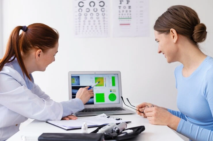
Laser iridotomy, also known as laser peripheral iridotomy (LPI), is a commonly performed laser procedure for people with narrow angles or certain types of angle-closure glaucoma. The treatment creates a tiny opening in the peripheral iris to provide an alternative pathway for aqueous fluid and help reduce the risk of sudden increases in eye pressure.

Although the procedure is generally quick and performed on an outpatient basis, recovery still requires appropriate aftercare. In this guide, we explain laser iridotomy recovery time, what you may experience during the first 24–48 hours, what you should and should not do, possible side effects, exercise restrictions, and when to contact your eye specialist.

What Is Laser Iridotomy?

Laser iridotomy is a laser treatment used primarily for eyes with narrow angles or those at risk of angle closure. During the procedure, an ophthalmologist uses a laser to create a very small opening in the iris. This opening helps aqueous fluid move more freely and can reduce the risk of an acute rise in intraocular pressure.

Because the treatment is performed with numbing eye drops and does not normally require stitches or general anaesthesia, patients can usually return home after a short period of observation.

Laser Iridotomy Procedure: What Happens?

Before the procedure, your ophthalmologist may use eye drops to prepare the eye and make the treatment more comfortable. The laser is then carefully directed at the peripheral iris to create the small opening.

The procedure itself is generally brief. After treatment, your eye pressure may be checked before you leave. Your doctor may also prescribe anti-inflammatory or pressure-lowering eye drops depending on your individual condition.

Because vision can be temporarily blurred after the procedure, arranging transportation home is advisable if your ophthalmologist recommends that you avoid driving.

What Happens Immediately After Laser Iridotomy?

The first few hours are an important part of laser iridotomy aftercare. Some patients notice temporary changes in the treated eye while it adjusts to the procedure and the medications used around treatment.

You May Experience:

  • Blurred vision: Vision may remain slightly blurred for a short period because of the eye drops or treatment.
  • Mild stinging or irritation: A temporary burning or gritty sensation may occur.
  • Light sensitivity: Bright lights may feel uncomfortable, particularly during the first few hours.
  • Mild headache or eye ache: Some patients may experience temporary discomfort.
  • Glare, halos, or a shadow: Temporary visual phenomena can occur as the eye adjusts.
  • Eye redness or watering: Mild irritation and watering may occur after treatment.

Your ophthalmologist may check your intraocular pressure after the procedure to make sure it has remained within a safe range. Follow the monitoring and medication instructions provided by your eye specialist.

Laser Iridotomy Recovery Time: How Long Does It Take?

For many patients, the most noticeable discomfort improves within the first day or two. Mild blurred vision, irritation, light sensitivity, or visual disturbances may sometimes continue for several days.

However, laser peripheral iridotomy recovery time varies from person to person. Your recovery may depend on your eye condition, whether one or both eyes were treated, your response to the laser, and any other glaucoma treatment you are receiving.

Important: Do not judge recovery only by how comfortable your eye feels. Attend the follow-up appointments recommended by your ophthalmologist so that eye pressure and the treated area can be assessed.

Laser Iridotomy Aftercare: What Should You Do?

Following your doctor’s instructions is the most important part of laser peripheral iridotomy recovery. Practical aftercare may include:

  • Use prescribed eye drops: Follow the exact dose and schedule provided by your ophthalmologist.
  • Give your eyes some rest: If your eyes feel tired or uncomfortable, reduce prolonged reading and screen use during the first day.
  • Wear sunglasses: Sunglasses can make outdoor light more comfortable when your eyes are sensitive.
  • Avoid rubbing your eyes: Keep your hands away from the treated eye unless instructed otherwise.
  • Keep follow-up appointments: Eye-pressure monitoring is an important part of post-procedure care.
  • Stay hydrated and maintain your normal healthy diet: Continue your usual balanced meals and adequate fluid intake unless your doctor has advised otherwise.

What Can’t You Do After an Iridotomy?

Many patients ask what they should avoid after laser treatment. Your ophthalmologist’s instructions should always take priority, but common precautions may include:

  • Do not rub or press the eye.
  • Do not drive until your vision has stabilised and you have been advised that driving is safe.
  • Avoid strenuous exercise immediately after treatment unless your ophthalmologist says it is appropriate.
  • Avoid heavy lifting or intense physical exertion during the initial recovery period if your doctor has advised restriction.
  • Avoid eye makeup while the eye is irritated or until your doctor says it is safe to resume.
  • Do not stop prescribed eye drops on your own.

Can I Exercise After Laser Iridotomy?

This is one of the most common questions about laser iridotomy recovery. Light activities such as gentle walking may be suitable for many patients, but strenuous exercise, heavy lifting, or intense workouts may need to be avoided temporarily.

The exact time when you can return to gym workouts, yoga, running, or other physical activities depends on your individual situation. Ask your ophthalmologist when it is safe to resume your normal exercise routine.

Common Laser Iridotomy Side Effects

Most post-procedure symptoms are temporary, but knowing the difference between expected discomfort and warning signs is important.

Mild and Temporary Symptoms

  • Mild eye redness
  • Temporary irritation or watering
  • Light sensitivity
  • Mild headache or eye ache
  • Slightly blurred vision
  • Glare or halos around lights

These symptoms generally improve as the eye settles after treatment. If symptoms are worsening, severe, or not improving as expected, contact your ophthalmologist.

Laser Iridotomy Risks: When Should You Contact Your Doctor?

Laser iridotomy is generally considered a safe procedure, but complications can occur. Contact your eye specialist promptly if you experience symptoms that are severe, worsening, or significantly different from what you were told to expect.

Seek Prompt Medical Attention If You Experience:

  • Severe or increasing eye pain
  • Sudden or significant reduction in vision
  • Marked redness or swelling
  • Eye discharge
  • Persistent or worsening halos or flashing lights
  • A sudden increase in floaters or black spots
  • Any symptom that causes significant concern after the procedure

Do not wait for symptoms to become severe if you are concerned about your vision. Prompt assessment can help identify and manage potential complications.

After Laser Iridotomy: What Happens Next?

Laser iridotomy is an important treatment for eyes with narrow angles, but follow-up care remains important. Your ophthalmologist may monitor your eye pressure and examine the treated area to determine how well the eye is responding.

The follow-up schedule varies depending on your diagnosis and individual risk factors. Some patients may require additional glaucoma treatment or ongoing monitoring even after a successful iridotomy.

Does Laser Iridotomy Hurt?

The eye is usually numbed with drops before the laser treatment. During the procedure, patients may notice pressure, a brief stinging sensation, or flashes of light rather than significant pain.

Some mild soreness, irritation, or headache can occur afterward. If pain becomes severe or continues to worsen, contact your ophthalmologist rather than assuming it is part of normal recovery.

Can You Wash Your Face After Laser Iridotomy?

Follow your ophthalmologist’s specific instructions regarding washing your face and caring for the treated eye. During the early recovery period, be gentle around the eye and avoid rubbing or pressing on it.

Can You Work or Use Screens After Laser Iridotomy?

Many patients can return to normal daily activities relatively quickly, but temporary blurred vision or light sensitivity may make reading, computer work, or prolonged screen use uncomfortable.

If your eyes feel strained, take regular breaks and reduce screen exposure during the first day. Do not resume activities such as driving until your vision is sufficiently clear and you have been advised that it is safe.

Laser Iridotomy Recovery: Important Tips for a Smooth Healing Process

  • Follow your prescribed eye-drop schedule.
  • Attend all recommended follow-up appointments.
  • Protect your eyes from uncomfortable bright light.
  • Avoid rubbing or pressing the treated eye.
  • Give yourself time to rest if vision is temporarily blurred.
  • Ask your ophthalmologist before returning to strenuous exercise.
  • Contact your doctor if symptoms become severe or worsen.

FAQs About Laser Iridotomy Recovery

1. How long does it take to recover from laser iridotomy?

Many patients experience improvement within 24–48 hours, although mild visual disturbances or irritation may last longer. Recovery time varies between individuals.

2. How long should I rest after a laser iridotomy?

Because laser iridotomy is generally an outpatient procedure, prolonged bed rest is usually not required. However, you may need to take it easy for the rest of the day and avoid strenuous activities according to your ophthalmologist’s advice.

3. Can I exercise after laser iridotomy?

Gentle activity may be acceptable for many patients, but strenuous exercise and heavy lifting may need to be postponed temporarily. Your eye specialist can give you a personalised timeline.

4. Will I still need glaucoma medication after laser iridotomy?

Possibly. Laser iridotomy addresses the anatomical risk associated with narrow angles, but some patients may still require medication or additional glaucoma management. Never stop glaucoma medication without medical advice.

5. Can laser iridotomy be performed on both eyes?

Depending on the patient’s eye anatomy and risk of angle closure, an ophthalmologist may recommend treatment for one or both eyes. Your specialist will determine the appropriate treatment plan.

6. Are glare and halos normal after laser iridotomy?

Temporary glare, halos, or other visual disturbances can occur after treatment. If they are persistent, worsening, or accompanied by pain or significant vision changes, contact your ophthalmologist.

7. Does laser iridotomy hurt?

Numbing eye drops are used during the procedure, so significant pain is not usually expected. Some patients may experience brief discomfort or mild soreness afterward.

8. What should I avoid after an iridotomy?

Avoid rubbing or pressing the eye, driving while your vision is blurred, and strenuous activities until your ophthalmologist confirms that they are safe. Follow all individual post-treatment instructions.

Final Takeaways: Laser Peripheral Iridotomy Recovery

Laser peripheral iridotomy recovery is usually straightforward, but temporary blurred vision, light sensitivity, irritation, headache, or glare may occur during the early recovery period. Most mild symptoms improve as the eye settles.

The best approach is to follow your ophthalmologist’s aftercare instructions, use prescribed eye drops correctly, avoid rubbing the eye, and attend recommended follow-up visits. If you experience severe pain, sudden vision changes, marked redness, discharge, or other concerning symptoms, seek medical attention promptly.

If you are considering laser iridotomy for narrow angles or need expert evaluation for glaucoma, consult an experienced ophthalmologist. A proper examination can help determine whether laser treatment is appropriate for your eyes and what recovery plan you should follow.